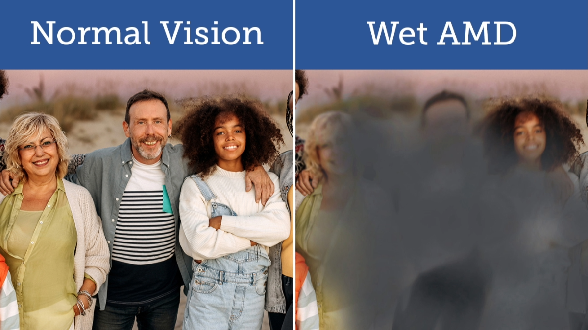
Wet age-related macular degeneration (AMD) can cause rapid vision loss. Learn how recent advances in treating wet AMD have decreased the rate of vision loss, and about research that may eliminate the need for injections.
What is Wet Age-Related Macular Degeneration?
Wet AMD occurs when new blood vessels grow into the retina. These vessels leak and bleed, eventually damaging the macula (the center of the retina) unless the affected eye is treated.
Progress in Preventing Vision Loss in AMD
Prior to 2005, most patients with wet AMD would lose much of their central vision over the course of a few months to years, depending on the severity. The disease would often progress until scar tissue replaced the vision cells in the macula, and patients wound up losing their central vision, keeping only the peripheral vision, which provides an acuity of 20/200 or less.
In 2005, the first of the new drugs that inhibit vascular endothelial growth factor (VEGF) became available and began providing remarkable vision protection. VEGF promotes the growth and leakage of abnormal vessels in the retinas of patients with wet AMD. Drugs that block VEGF typically work for 4-12 weeks, and then need to be injected again. These drugs include brolucizumab (Beovu®), aflibercept (Eylea®), ranibizumab (Lucentis®), and bevacizumab (Avastin®). Clinical trials indicate that in some patients, Eylea can last as long as 8 weeks and Beovu as long as 12 weeks.
The Eye Injection for Wet AMD
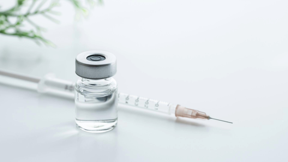
VEGF drugs are administered by injection into the vitreous jelly in the center of the eye. The eye is numbed with drops, cleaned, and the injection, which is only moderately uncomfortable for most patients and takes only a few seconds, is done through a tiny needle. Some ophthalmologists prefer to give the injections on a regular schedule, while others will base the re-injection decision on the results of a special type of retinal photograph called optical coherence tomography (OCT).
Eye Injection Risks
The injection risks are low: about a one in a thousand chance of retinal detachment or bacterial infection in the eye (endophthalmitis). Beovu causes inflammation in about 4 percent of eyes, and this has been associated with diminished vision caused by inflammation of blood vessels (occlusive vasculitis) in 11 out of 46,000 injections so far in the US.
How will Vision Improve After the Eye Injections?
After the first few injections of an anti-VEGF drug, on average, vision improves by a couple of lines on the eye chart. Over the course of a few years, the vision tends to drop a bit, down to the pre-treatment level. It can get even worse, especially in patients who opt to have only very infrequent, or no follow-up injections. Some patients find it challenging to get frequent follow-up injections for a variety of reasons, including difficulty with transportation (some can no longer drive), other simultaneous health issues that need urgent attention, or cost. Avastin, which is used off-label, is the lowest cost drug.
The Future of Wet AMD Treatment
Many patients have asked me when longer-lasting drugs will become available, and some are in clinical trials. One is an implanted, inside-the-eye, storage device for Lucentis, which in some patients can last more than a year between refills. Also, two types of gene therapy, one injected into the vitreous, and the other injected under the retina, are now being tested, and early results suggest they may decrease or eliminate the need for further injections.
About BrightFocus Foundation
BrightFocus Foundation is a premier global nonprofit funder of research to defeat Alzheimer’s, macular degeneration, and glaucoma. Since its inception more than 50 years ago, BrightFocus and its flagship research programs—Alzheimer’s Disease Research, Macular Degeneration Research, and National Glaucoma Research—has awarded more than $300 million in research grants to scientists around the world, catalyzing thousands of scientific breakthroughs, life-enhancing treatments, and diagnostic tools. We also share the latest research findings, expert information, and resources to empower the millions impacted by these devastating diseases. Learn more at brightfocus.org.
Disclaimer: The information provided here is a public service of BrightFocus Foundation and is not intended to constitute medical advice. Please consult your physician for personalized medical, dietary, and/or exercise advice. Any medications or supplements should only be taken under medical supervision. BrightFocus Foundation does not endorse any medical products or therapies.
- Prevention
- Treatments
- Wet AMD


