Can Vision Loss Be Reversed? Exploring Stem Cells and AMD
Featuring
Kapil Bharti, PhD
Senior Investigator and Director, Intramural Research Program
National Eye Institute, National Institutes of Health
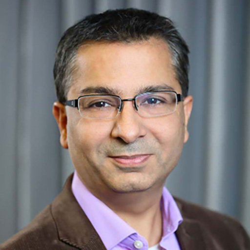

Kapil Bharti, PhD
Senior Investigator and Director, Intramural Research Program
National Eye Institute, National Institutes of Health

Guest speaker Kapil Bharti, PhD discusses a new treatment for macular degeneration that is currently being studied using stem cell therapy to restore lost vision.
Dr. Kapil Bharti’s lab at the National Eye Institute recently started the first U.S. phase I/IIa trial to test autologous iPSC-derived RPE patch in AMD patients. Currently, he is co-developing a dual RPE/photoreceptor cell therapy with Opsis Therapeutics. He has given several keynote lectures and won several awards for his revolutionary work on developing ocular cell-therapies. He serves on the advisory board of several companies and patient-advocacy groups. His current work as a Senior Investigator at NEI involves understanding the mechanism of retinal degenerative diseases using induced pluripotent stem cell derived eye cells and tissues and developing cell-based and drug-based therapies for such diseases. He is the Director of the NEI Intramural Research Program where he oversees 21 research labs and 6 core facilities.
Dr. Bharti is also a Macular Degeneration Research grantee. Learn more about his funded work here: Engineered Eye Tissue Models to Analyze Mechanisms of Age-related Vision Loss
Download English Transcript PDF
DR. DIANE BOVENKAMP: Hello, and welcome. My name is Dr. Diane Bovenkamp, Vice President, Scientific Affairs, at BrightFocus Foundation. I’m pleased to be here with you for today’s macular degeneration Chat, “Can Vision Loss Be Reversed? Exploring Stem Cells and AMD.” This Chat is brought to you today by BrightFocus Foundation. Macular degeneration research is one of our programs here at BrightFocus. We fund exceptional scientific research worldwide to defeat Alzheimer’s disease, macular degeneration, and glaucoma, and we provide expert information on these heartbreaking diseases. Just sit back and relax and enjoy our wonderful scientific discussion. I know I’m excited, and I’m totally pleased to introduce today’s guest, Dr. Kapil Bharti, who’s the Senior Investigator and Director of the Intramural Research Program at the National Eye Institute of the National Institutes of Health—some of you may have heard NIH. Dr. Bharti’s lab at the NEI recently started the first U.S. Phase 1/2A trial to test autologous iPSC-derived RPE patch in AMD patients. And there’s a lot of alphabet soup in there, but by the end of this talk, you’ll know what all of those acronyms mean. Currently, he is co-developing a dual RPE photoreceptor cell therapy with Opsis Therapeutics. He has given several keynote lectures and won several awards for his revolutionary work on developing ocular cell therapies. His current work as a senior investigator at NEI involves understanding the mechanism of retinal degenerative diseases using induced pluripotent stem cells—that’s the iPSCs—derived by cells and tissues and developing cell-based and drug-based therapies for such diseases. Dr. Bharti, thank you so much for joining us today.
DR. KAPIL BHARTI: Oh, Diane, it’s my absolute pleasure. Thank you very much for having me here. And before we begin, I actually do want to applaud you and the BrightFocus Foundation for supporting really amazing basic and translational research in the macular degeneration field, and for organizing events like you’re organizing today to educate the community on macular degeneration, its disease pathology, and the treatments that many of us are working on.
DR. DIANE BOVENKAMP: It’s our absolute pleasure to do that, and it’s just wonderful to be a partner with you as we try and push forward to help all affected families, so thank you so much. Before we jump into our exciting conversation exploring stem cell research, I did want to mention that the contents of this Chat are intended for informational and educational purposes only and not for the purpose of rendering medical advice. Please consult your physician for personalized medical advice. Always seek the advice of a physician or other qualified health care provider with any questions regarding a medical condition, and especially before beginning any new treatments or clinical trials. Sorry, had to do that legal disclaimer, especially since you work for the government and we’re also a nonprofit. In summary, go to your doctor if you have any questions. Anyway, I think that a lot of people are interested in … they hear “stem cell this” and “stem cell that.” Can you just explain to us: What is a stem cell?
DR. KAPIL BHARTI: So, stem cell. The definition of a stem cell is very simple. It’s a cell that can make more copies of itself under certain conditions, and other different conditions, it can make other cell types. I think the most classical example I like to give is the bone marrow stem cells, the cells that make our blood. We all know that blood cells replenish every 3 months or so, and that’s because we have blood stem cells in our bone marrow and in our spleen, and these cells keep making, keep replicating, and keep maintaining themselves as blood stem cells, but every 3 months, they keep making new copies of blood cells for our body. And that’s a very focused stem cell, or what you call a cell that has very limited potential—it cannot make any other cell type beyond blood cells. But then stem cells like iPS cells that you mentioned earlier—induced pluripotent stem cells that can be made from any adult tissue, any adult cell—it has the potential to make any cell type of the body. That’s why it’s called “pluripotent.” In a dish, we can keep the cell forever as a stem cell, or we use it to make cells of the eye, cells of the heart, cells of the lung, the liver, the brain, you name it. So, that’s a stem cell. Multipotent, if it’s a blood stem cell, it can only make limited cell types. Pluripotent, if it’s an iPS cell, it can make any cell type of the body.
DR. DIANE BOVENKAMP: And these have to do with what tissue they’re in, what environment they’re in, and all of the proteins and chemicals these stem cells are bathing in, right? It kind of defines the identity it ultimately becomes.
DR. KAPIL BHARTI: It depends on what kind of stem cell it is, Diane. If it’s a multipotent stem cell, like the stem cell that makes the blood, that, of course, is defined by the environment it is in—that is a bone marrow, that is a spleen—and it can only make blood stem cells. We all go to get haircuts; that’s because we have stem cells in those hair shafts inside our skin that keep making the hair protein, right? So, that stem cell will only make hair cells, right? We have stem cells in our muscles. We all know that if we exercise, our muscles regenerate, they become bigger, they become stronger. And that’s because we have stem cells in our muscles, but those stem cells will only make muscles, whereas iPS cells are actually an artificial class of stem cells. They are not present in our body. We induce them in a dish from patients or from anybody’s skin cells or blood cells. We just need a handful of cells. I would need half a cc of blood, or even less than that, to make iPS cells from anyone we can in the lab. And these cells, for practical purposes, are very identical to—almost identical, I would say—to the embryonic stem cell, the infamous stem cell that people have talked about for a while and have a lot of political issues associated with that. Embryonic stem cells are present in a blastocyst in an embryo. They make our entire body. We’ve all seen that or heard about that; iPS cells are identical to embryonic stem cells in a way that these cells can also make all of the cells of the body, but all the things that we do with them are happening in a dish—they are not present inside of a body.
DR. DIANE BOVENKAMP: Great. So, that’s a good point. You talked about hair, and you talked about spleen and muscle. Would you explain a bit more about your specialty, which is retinal pigment epithelium, or RPE, cells. How do these iPSC cells become RPE or other cell types to prepare for future transplantation stem cell therapies?
DR. KAPIL BHARTI: Yeah, good question. So, an RPE cell—that’s an acronym, retinal pigment epithelium, as you said—this is a cell type that is present behind the retina. We should think about it this way: People who have been here long enough know that we used to use polarized cameras—polaroid cameras—in the past. We would have always a black screen in the back and a light-sensitive filament in the front, and the light-sensitive screen is what captured the light, and that’s where the image was formed. Very similar to that, the retina is the light-sensitive part that perceives light and sends signals to the brain so we all can see, whereas the retinal pigment epithelium is a little bit of a dark screen in the back that protects the light from hitting the rest of our brain and also shows us direction of the light. So, it has that function, but has many other functions. It nourishes the retina throughout its life. It helps protect the retina from injuries and from damage due to inflammation and things like that. So, this tissue, the RPE, is very important tissue for us to be able to functionally see on an everyday basis. If RPE cells don’t function properly, our photoreceptors switch out the light-sensitive cells in the retina, they start to die, and, of course, with diseases like macular degeneration, that’s what happens, and patients would go blind. Unlike the muscle or the blood or the hair, RPE cells do not have any stem cells in them, or not widely known stem cells that can keep regenerating new RPE cells. So, once an RPE cell stops functioning properly and eventually starts to die, there’s no way for the body to replace them. And that’s the reason, Diane, that patients go blind because when RPE cells die, as I said, the photoreceptors will die.
So, what surgeons tried for a long time was that when they saw that in macular degeneration patients, it’s only cells in the center of the eye, literally almost a 5 millimeter area, which is like one-fifth of an inch, is the area that dies off. But that area, the center part of the eye, called the macula—hence the disease macular degeneration—that area is the most important part for our vision. So, what surgeons would do is say, “Okay, if the RPE cells are dead here in this area, what if you bring fresh RPE cells from the periphery of the same eye and put them in the center? Would this support the photoreceptors and perhaps protect the photoreceptors from dying and maybe stop the disease from progressing further?” You can imagine it’s a complicated surgery because they have to cut out one part of the eye, put it to the other part of the eye. But in some number of cases where the surgeons were successful in doing that surgery correctly, they saw that the RPE cells that were transplanted in this new area, they survived for years to come and they supported the photoreceptors for years to come. So, that, for us, provided the proof of concept that if we can make these RPE cells from patients on stem cells—patients on iPS cells that we talked about a few minutes ago—in a dish, well characterize them, and then bring them at the right place, which is the macula, at the right time of the disease, we have a fighting chance of stopping this disease from progressing further.
That’s the whole approach that we have taken from iPS now to making the RPE cells in a dish. We use classical developmental biology that we have learned that BrightFocus has supported over the years, how RPE cells have made in an embryo as an embryo forms. As the eye forms, how do RPE cells differentiate? And this work has been done in many different species—mouse, rat, chick—so many different areas where RPE development has been studied. We took all of that knowledge, and we transferred that into making the best possible way, the fastest possible way of RPE cells from iPS cells in a dish, and that protocol worked great. We can now, within 10 weeks, make from patients’ own iPS cells a fully mature, pure culture of RPE transplant that we can bring back to patients’ eyes.
DR. DIANE BOVENKAMP: That is just so amazing how that’s what science is. Scientists are really generous, they publish the work, and then so whatever work you’re doing, you’re building upon the shoulders of past giants, right? What you’re doing today is a culmination of many years’ work, and so, can you explain … I think that maybe some listeners are saying, “Well, why do you need to just get cells from the same person?” and I think it has to do with transplantation rejection, right? Am I right in that? Instead of just getting just a generic iPS cell, you want to get a cell from the same person, so it’ll stick?
DR. KAPIL BHARTI: That is absolutely correct, Diane. Because that example, that surgical example I gave you, there these were patients’ own RPE cells from the same eye even, right? And they transplanted, they engrafted very well, and that’s because the immune system did not see them as foreign cells. They said, “Oh, that’s our own cells!” But, of course, the problem with that procedure was we’re still cutting out a part of the eye, and these were still old cells, they were still diseased, and so there were a lot of issues, and the surgery was very complicated. But in this case at least took that knowledge, and, like you said, for making RPE cells, it is literally the last 30 years of work that we put together from biggest giants in developmental biology field to put together that protocol, but for making this transplant what we call an autologous transplant as compared to an allogeneic. Allogeneic would be somebody else’s cells in you or in your eyes; autologous is your own cells in your eyes. The reason is, like you said, the immune system would not see them as foreign cells, it would not react against them, and they would have a very high chance of engrafting and immediately start helping the retina and the photoreceptors and start nourishing it so that the patient can see again.
DR. DIANE BOVENKAMP: Yeah, great. So, I guess, what is the advantage of using stem cell–derived therapy for macular degeneration? Because we know if you have wet AMD, you have the shots to try and reduce the bleeding, reduce the blood that can collect, and now, just in the last year or so, there’s some treatments for dry AMD based on C3 and C5 inflammation proteins. You know, that’s a topic of another Chat, so listeners, go to one of those. But yeah, so what’s the advantage of using the stem cell therapy instead of other treatments?
DR. KAPIL BHARTI: Yeah, good point, Diane. So, both of these treatments that you mentioned for wet AMD, which has been around for many years now, and the dry AMD treatment that just came out this year—both of these are treatments that slow down disease progression and manage the disease. They do not really cure the cells that have been gone. They do not regenerate the cells that have already been gone. And in both cases, first of all, you have to get almost a monthly injection—sometimes bimonthly—but it’s an injection in the eye. It’s an extremely uncomfortable thing to have, and you have to have those for the rest of your life. Whereas the stem cell therapy, and like I said earlier, because the RPE cells and the photoreceptor cells do not have any stem cells in them, once they are dead, they are dead. The injections are not going to bring the dead cells back; the injections of anti-VEGF or anticomplement inhibitors are not going to bring the dead cells back, whereas the stem cell–derived transplant is going regenerate those cells. We are bringing fresh, young, new cells in the eye with the idea that these cells will live for the next several years—it’s a one-time surgery, and hopefully the cells will stay for the rest of the life of the patient. They will not have to get a monthly injection. So, that’s a big advantage. So, if and when it works, it’s a curative therapy as opposed to a treatment.
DR. DIANE BOVENKAMP: Great. It will help patients who might have become refractory or those treatments don’t work for them sometimes, and/or the disease has progressed enough, as you said, that the cells have died, so this is really amazing. At what stage of age-related macular degeneration do you think that stem cell–derived therapy has to be administered?
DR. KAPIL BHARTI: Yeah, again, a good point. Stem cell–derived therapy should be done ideally at the time when RPE cells are dying so that we can bring RPE cells in before too many photoreceptors have died, because once the photoreceptors are dead, RPE cells are not going to bring new photoreceptors, so then we have to think about double RPE photoreceptor therapy, which maybe we’ll talk about later, but many of us are working on now. But this approach of RPE transplant is going to happen at a stage when disease has progressed to the stage that RPE cells are starting to die. So, you will bring in transplant the RPE cells and have them protect the photoreceptors. Now, if you think about late-stage dry AMD, which is the geographic atrophy stage of AMD, that lesion starts off small, and it keeps growing every year about 20 to 30 percent, right? And it grows because at the edges of that lesion, RPE cells continue to die. So, our first approach is going to be to transplant this patch in that lesion, in those edges area, and stop it from growing. And then, as we get more and more comfortable that the technology is working, the drug is working, we start transplanting them earlier and earlier to the point that we don’t even have much significant photoreceptor cell that … so the patient’s vision is not significantly gone by the time we have done the transplant.
DR. DIANE BOVENKAMP: Yeah, great. Yeah, it is still in progress, right? And you can tell us about the trials that are going on in a little bit, but I know that there’s a lot of benefits and hope associated with this stem cell therapy, but, you know, we always want to look at: Are there any risks associated with this?
DR. KAPIL BHARTI: Risk associated with stem cell therapy. So, you know, we have to think about what this drug is. Unlike a pill that we all are used to taking or an eyedrop that many of us take, you know, at the time of allergies and whatnot, stem cell therapy is a living drug. These are living cells we are transplanting; these are patients’ own tissues we are transplanting back in their eyes. And these cells, there’s a lot that we know about them, but we don’t know what we don’t know, right? So, we have to take that approach and cautiously work with this technology, like any other drug that we work with, but like I said, this drug is a magnitude more complex because it is a living drug. We test it in many ways in the lab, we test it in animals before we transplant that in patients, but it’s going to react to the patient’s own body, to the patient’s own tissues, their own inflammation, their own everyday lifestyle. This drug will react and may behave differently. So, there is a lot of unknowns, and as we are learning and as we are doing more and more transplants, we’re learning more about this. And that is one of the biggest things, I would say, is concerns with stem cell therapies is that there’s a lot that we don’t know that we don’t know.
Then, of course, because these are stem cells, as when we started the call, we talked about that these stem cells can become any other cell type of the body as well, right? So, we have to make sure that we are not transplanting—and I want to make it absolutely clear to our audience—is that we are not transplanting stem cells. We are transplanting stem cell–derived eye tissue that we make in a dish, and we go through absolute lengths to make sure that there’s not a single stem cell left in that final tissue. FDA does not allow us to go to patients until we demonstrate in five different ways that there’s no stem cells left in our final tissue, because if there was one, it has a chance that it can make any other cell type of the body. But since we go to lengths to purify that, to show that doesn’t happen, we are fairly confident that we are transplanting pure eye tissue in patients’ eyes.
DR. DIANE BOVENKAMP: Yeah, that’s a really good point. I mean, I come from originally, I was in the cancer research field. And the definition of a cancer cell is uncontrolled growth. So, I guess you want to make sure that the stem cells are under controlled growth so it doesn’t turn cancerous as well, right? I think that’s one of the risks.
DR. KAPIL BHARTI: Yup and that’s the reason when we convert the stem cells in a dish into RPE cells, we ensure that there is no stem cell left. And RPE cells, once they’re fully mature, they’re not going to divide anymore. So, those are the reasons we have worked on this. We—and when I say “we,” the whole group or the whole field who is working in this space—have ensured that we are going to only transplant the final tissue, not the original stem cells, right? So, this tissue will not have potential to continue dividing; this tissue will only go in to help the retina, to help protect the retina, to nourish the retina. These cells will not have any proliferation potential left when they are transplanted in the eye.
DR. DIANE BOVENKAMP: Great, and yeah, and I think that we don’t want to cause undue panic with people. We just want people to know what are the risks and benefits—before you get any treatment you want to know the benefits. And I think I was going to ask this later, but I think this is probably a good time. The process that you go through and other groups have gone through the FDA has tight regulations, and there’s a lot of ethics involved, and as you said, you want to make sure that nothing is remaining as a stem cell that can change. But here are some stem cell clinics you hear of on the news or some people say, “Oh! Go to Mexico or whatever and pay us X number of dollars, and we’ll put stem cells in your eye and get your vision back.” Do you have anything to say about that? Maybe as a public service or give us more information about that in case some of the listeners have heard of those?
DR. KAPIL BHARTI: Diane, this, I think, is one of the most important pieces of advice we could give our audience today. When you say “stem cell clinics,” I would put them under quotations, because these are not true centers who are trying to develop stem cell therapies. These are the places who are abusing patients’ desperation to get their vision back to transplanting something that is not even a real cell type that should be there. The cases that I’ve heard about is they would take out some fat stem cells from the hip or from the fat in the body and inject it in the eye. While we started this call by talking about specialized stem cells versus iPS cells, which behave differently, and we talked about that blood cells—blood stem cells—will only make blood cells, the same way fat stem cells will only make fat tissue. So, imagine you inject fat stem cells in the eye and they end up making fat in the eye—and that actually has happened. There have been cases in which some patients were transplanted with such cells in Florida, and three people actually went completely blind because of that procedure.
So, my word of advice is for patients to please, please be extremely cautious whenever they hear something about this. Please ask them if this procedure has been approved by the FDA for a Phase 1/2 study—that’s the first thing. And if it hasn’t been approved, then it is not a real experimental drug. It is somebody who’s trying to make money off it. And the second thing: Anybody who is doing a legitimate trial that has been a clinical trial that has been cleared by the FDA and by academic agencies or academic centers, they will not charge you to participate in that study, whereas most of these “stem cell clinics” will charge you anywhere between $25,000 to $50,000 for those procedures. So, please be extremely careful. NIH has on their website a lot of information about differences between legitimate and so-called snake-oil in the stem cell field. FDA has that information on their website. [International Society for Stem Cell Research], ISSCR, has that information on their website. Please read the information, please educate yourself, and please don’t be harmed by these snake-oils that are out there.
DR. DIANE BOVENKAMP: Great, and you know what, I think maybe what we’ll do is we can get all of those links to extra information from you after and maybe post it at the end of the transcript when it’s made just so people will know where to go.
DR. KAPIL BHARTI: Absolutely.
DR. DIANE BOVENKAMP: Thank you so much, so much, because we just want everybody to protect themselves. Before we go on to talk about more specifically about your research and your team’s research, I see that there is one question that came in from someone that says, “How do you know when the RPE cells are starting to die?” And then they said, “Do you use OCT or another method?”
DR. KAPIL BHARTI: Great question. Yes, OCT is one of the most commonly used technologies. OCT stands for optical coherence tomography. It’s a technique where they shine light into the eye, and they get reflectance back from the retina, and from there the technology can tell how different layers of the retina, as well as the RPE, are structurally intact or not. There are other techniques that our physician scientists and physicians use to look at if RPE cells are dying. Another structural feature is called adaptive optics, where they can look at single-cell resolution details of the cells and see if the cells are alive or dead. The functional measures of the vision that they can use is called microperimetry, dark adaptation. These are all different techniques that tell structurally and functionally if the retina and the RPE are intact, and from those, from very early on, they can start to tell you if RPE cells are dying and will have an immediate impact actually on your vision.
DR. DIANE BOVENKAMP: Great, thank you. Thank you, and I think we might have some information on our website people can go and look up, or we can put links to what all of those devices are so they can print it out and bring it to their doc. Okay so, as I said before, I am extremely proud that BrightFocus is funding you, and we funded you in 2020, and also we’re currently funding one of your postdocs, of which you’re a mentor on them. But the grant in 2020 you received for Macular Degeneration Research is you’re working with something that’s really cool that some people might have heard of. It’s like 3D bioprinted human tissue models, so kind of like 3D printing, but it’s not just like spurting out ink, right, or molten plastic, if you go to the library and you use CAD and whatever, and you create these little 3D things. You took this one step further. So, you’re bringing in engineering, and you’re bioprinting human tissue models to clarify the role of retinal blood vessels in the retina and about giving more information about macular degeneration itself. This is so fascinating. Can you tell us a little bit more about this research?
DR. KAPIL BHARTI: Diane, first of all I have to say you summarized it really well. Thank you very much. So, essentially you are absolutely right. We are bringing engineering and biology together, and the idea comes from what we talked about earlier in the call, is that there are two types—two advanced stages of AMD. One is the dry AMD that we are trying to treat with these RPE transplants, in which case we talked about the RPE cells die, but we also talked about that RPE cells are nourishing the photoreceptors and the retina. They’re bringing nutrients from the blood supply that is present underneath the RPE and taking the photoreceptors, and they take all the metabolites from the photoreceptors, they take it back to the blood supply. That’s the one important function RPE cells perform. And in dry AMD, as RPE cells die, the blood supply—also underneath—the blood supply starts to die, and that whole relationship between RPE cells’ function, nourishing the retina, how they take the nutrients from the blood supply, bring it to the retina, and why the blood supply dies once the RPE cells die is not clear. And then the opposite thing happens in the other advanced form of AMD, called the wet AMD, where the blood supply grows too much, and then what I like to call it is grows in the wrong direction, or the Z direction, and it penetrates through the RPE, ruptures the RPE barriers, and leaks fluid into the retina and blood into the retina, and then, of course, leads to blindness. So again, then it’s not clear why, in the case of wet AMD, the capillaries divide too much, they proliferate too much, whereas in dry AMD, they die.
So, we thought we need to recreate this tissue in a dish so we can better understand the closed interaction, the symbiotic interaction that we think is there between the RPE and the blood supply. To achieve that, we combine the stem cell biology, the developmental biology that we have been talking about, that from iPS cells we can not only make the RPE cells, we can make cells of blood vessels, the endothelial cells, the pericytes, the fibroblasts. And then what we do is, by combining that whole thing with 3D bioprinting, which essentially is a little bit fascinating instrument that has two syringes attached on it which we can control in a very precise XYZ orientation. And in those syringes, we fill in cells mixed with hydrogels, and in this case, the cells that would make the capillaries—that would be the endothelial cells, pericytes, and fibroblasts. And then, we go around in our dish and we make the capillary network artificially with that structure, with that syringe, by moving it in a very precise way. And the cells, because they are coming from stem cells, they have a natural tendency, they know what to do. They start doing their job within literally 1 week. The endothelial start to make vessels and capillaries, and pericytes wrap themselves around and start supporting the lumen of them, and fibroblasts fill in the matrix in between, make the matrix so that the capillaries can stay stable. And then we … on the other side, we put a scaffolding between on the other side, which seed the RPE cells, again, made from iPS cells. And in about 5 weeks or so, the whole tissue matures. And it was really fascinating to see that we make genetic tissue, but when they start talking—the RPE cells and the capillaries start talking to each other—they start behaving like they would in the eye. They have same gene expression pattern, they have the same functionality. And then we tried to do what was the most difficult part—we tried to replicate the dry AMD phenotype and the wet AMD phenotype by different type of stressors in that dish.
So, I told you that in dry AMD, when RPE cells are not happy, capillaries die. We did the same experiment in a dish. We made RPE cells unhappy, and we saw that the capillaries died. And then we did the opposite experiments, where we caused hypoxia in RPE cells, which is known to secrete this chemical called VEGF, vascular endothelial growth factor, which is known to cause wet AMD. And guess what? The capillaries made wet AMD in a dish, which we could suppress by adding anti-VEGF antibodies. So, this gave us a lot of confidence that the tissue that we have recreated in a dish has a lot of clinical potential, has a lot of translational potential. It can not only be used to better study how disease progresses, how it initiates, it can be used to discover new drugs that will slow down disease progression, that will slow down cell death, that will slow down capillary cell death. And I have to say thanks a lot of thanks to BrightFocus Foundation who funded a lot of this work early on. Very early on you guys saw the potential of this work before we had published it and before we had completed it. And that led us to really complete a lot of the work that went into one of the manuscripts where we were trying to understand how these two tissues work together, and now we’re taking it to the next step where we will be making patient-specific tissues with different types of risk alleles and trying to understand how AMD progression happens in these patients.
So, that was one of the works that you funded, as you said, in 2020, but BrightFocus has also thankfully funded one of the fellows in my group, who, by the way, we just nominated him for the NEI Directors Rising Star Award, and he got that award. And he is! He is indeed our rising star. What he has done is he has combined artificial intelligence with image analysis to really better understand the different types of RPE cells that are present in human eyes and discovered that there are five different subpopulations of RPE cells that are present in different areas of the eye, and they have slightly different functions. And, in fact, if you look at diseases like macular degeneration versus another monogenic disease called choroideremia, different populations die.
So now, this gives us a handle how to study, specifically macular degeneration, in one specific subpopulation versus choroideremia in a different subpopulation and what Davide is doing, again thanks to funding from BrightFocus Foundation, is looking at why the cells in the macular, why the RPE cells in the macula in a patient’s eye die and not far away from it. So, that means he has an internal control, and since he can recreate both the central cells and the peripheral cells in a dish from iPS cells, he is able to compare the difference between them. He is able to compare the differential disease sensitivity to better understand why disease only happens in the center—not in the periphery. And then, I think that would be very important piece for us to perhaps even prevent disease from happening as opposed to treating it when its further gone and we have to bring the whole tissue made from stem cells. And Davide’s approach will hopefully help us bring new drugs for disease prevention as well. So, those are the two studies that I wanted to highlight. Both are funded by the BrightFocus Foundation, so thank you very much.
DR. DIANE BOVENKAMP: Oh my gosh, and thank you to all of the donors out there who are probably listening right now. Thank you for donating to us to help support this. And I think, and just to clarify, that is Dr. Davide Ortolan. We have info on the website, and you’re one mentor, and Dr. Ruchi Sharma is another mentor, too, so you can look that up. You really underlined the importance of funding basic research, because we still don’t even know a lot of the basic reasons why this disease starts. And the stem cell treatment is amazing, and we’ll talk a little bit more in just a second about, you were part of a big team that’s moving that forward, but like what you said, prevention is so much better. If we can try and figure out how to prevent the disease from even happening, then that would be amazing. But thank you so much. We’re just so honored to be able to fund your research. So, in the summer of 2022, last summer, you were part of a team that surgically implanted replacement tissues from patient-derived iPS cells, kind of like an iPS cell–derived RPE patch—this patch that you were talking about—to treat advanced dry AMD, this geographic atrophy that you were talking about. Can you tell us a little bit more about that procedure and what was done—and, of course, what you could publicly tell us about because if the clinical trial isn’t done yet, you can’t tell us any of that.
DR. KAPIL BHARTI: Yes, Diane, thank you for asking that question. So, this is really a culmination of almost 10 years of our work, trying to develop this treatment made from patients’ own stem cells, patients’ own iPS cells, as we have talked in the last half an hour. What we did was recreate the RPE monolayer in a dish on a scaffold that is biodegradable and degrades within a few weeks, and so some of it is outside, some of is in the eye, and the degradation products are not harmful to the eye. So, we were able to make a small piece—about 8 square millimeters, which covers a good part of the macula—and the procedure is obviously, as we have talked in the last few minutes, is that this kind of procedure will be applicable only to very late–stage patients where RPE cells are dead and we want to protect the photoreceptors by transplanting new RPE cells. So, those were the patients chosen for this procedure, and essentially, it’s a surgical procedure where we go from the side of the eye and make an incision in the retina and transplant the tissue under the retina, the RPE tissue under the retina—that’s where the RPE cells are naturally present anyways—and in the area where the RPE cells are gone because of the disease, the RPE cells have already died. And that procedure was done in the first patient last year. This was the first patient in the United States where such a procedure was done. I should say that a similar procedure has been done in Japan many years ago once, and that patient was … his eye has been stable and that patient in Japan was a wet AMD patient, and what I hear is that patient did not need any more injections of anti-VEGF therapy.
Similar different approaches, slightly different approaches are underway using allogeneic RPE patch. So, as we talked earlier, allogeneic means it’s somebody else’s cells, so these are primarily RPE cells made from embryonic stem cells. But a similar patch—in their case, put on a piece of plastic sheet—is made on a plastic membrane, and a trial has been underway in California, and a similar trial has been underway in London. So, many groups are moving forward. We’re all learning from each other, and the goal is really to bring this technology, bring this drug more as a common practice to everyone. We’re all at early stages. We’re all, as we have talked about earlier, walking this line cautiously to make sure that we are keeping patients safe and away from harm, but the hope is that one day we will make this available, most widely available, to as many patients as possible. The trial at NEI is still ongoing. It was unfortunate that the trial started in the middle of the pandemic or the start of the pandemic and we could not transplant anyone. Because of the pandemic, we all know that we had to shut down several clinical operations, but right now the trial has picked back again, and we are hoping to transplant more patients in the coming several months, and we will keep you posted as we go along with this journey.
DR. DIANE BOVENKAMP: What’s the projected timeframe to have everyone enrolled and treated and have their results ready to publish?
DR. KAPIL BHARTI: Good point. So, for a Phase 1/2A study, typically it’s anywhere between 2 to 5 years that the study lasts. For us, it went a little longer because of the pandemic, as I said, but we hope that in the next few years we will wrap up the study. We realized that our enrollment had been slower because of all those reasons that we talked, so we are trying to now open two additional clinical sites, which will be announced early next year, that will help us do this procedure, run this trial at three different sites. That will speed up the enrollment, and we hope to complete our enrollment in the next couple of years, and that’s when we hope to announce the results. Usually, the way any trial is done, any drug development requires different stages. Phase 1/2A is usually safety and visibility stage—that’s where we are. And then we will move on to what is called the Phase 2B, where we will have first signs of efficacy, and because the stem cell drugs are so complex, so complicated, and take so long, FDA allows them to get through a faster track of approval. So, that means if the Phase 2B results where we might be transplanting between around 60 to 80 patients, if those results look promising, if we see signs of efficacy, then already in Phase 3 it can be a registration trial. That means patients can register, there can be reimbursement from the Medicare for this kind of work, and it can be most widely available, as well. But you’re looking at all of this combined. You’re looking at anywhere between 5 to 8 year timeline until this is widely available.
DR. DIANE BOVENKAMP: Okay, so your trial is still going on. There’s other trials going on. And, of course, to be able to enroll in a trial you have to meet the enrollment criteria and yet not have any of the exclusion criteria, and each trial is different. So, just in case there are some people listening who might want to consider joining your trial or other trials, are there other active clinical trials and where can they find out about them and yet avoid those nasty in quotation “stem cell clinics” that aren’t FDA approved? How can people find a legitimate trial to enroll in, and what are they out there for AMD right now?
DR. KAPIL BHARTI: Yes, so, Diane, there are many trials for AMD that are ongoing, and all of those are listed at ClinicalTrials.gov—I repeat, ClinicalTrials.gov—where all of this information is available. You go in the simple search tab. If you look for NEI iPS RPE trials, you will find that information; similarly, you will find other trials. I have to warn, though, that the “stem cell clinics” also have their information on ClinicalTrials.gov, but you will see that on the second page there has to be a clear disclaimer—for instance, for our trial—that it has been cleared by the FDA for a Phase 1/2A study, and those clinics cannot put such a disclaimer because they haven’t gone through the FDA yet. So, please look out for that disclaimer. Ask the right questions to the study coordinators so that you are enrolling in a legitimate trial. And as you said, Diane, the early-stage trials have much harder, stricter eligibility criteria, but as we get more and more comfortable with the safety of the drug, the criteria get more and more relaxed, and a larger number of patients can participate in those trials.
DR. DIANE BOVENKAMP: Great, yeah. That’s really great, so to look on that second page to see if they were approved by FDA. There’s sometimes a code or something. And then also, they can probably just print that page out and bring it to a health care provider, too, to ask them about it. And I guess on that page too, there’s normally a telephone number that people can call to get more information, right?
DR. KAPIL BHARTI: That is correct. Yeah.
DR. DIANE BOVENKAMP: Okay, great. All right, anything else about the stem cell clinical trials before I go on? I mean, this has been just so fascinating.
DR. KAPIL BHARTI: I do want to put out a word of caution that sometimes when people hear the word “stem cell” they think there is a magic bullet here and it will cure and treat everything. I agree that stem cell–derived therapies do have curative potential, but just like any drug, it’s a lot of work in progress, and there’s a lot of iterative work on our side to make sure that it is done correctly. And often, we are going back from patients and changing some things and coming back to patients, so it may take time. And especially in early-stage patients, I don’t think we may see big signs of efficacy. We will have to wait until these drugs are perfected, until they reach the right stage of patients to really see their true potential of curative treatment. But I am hopeful, and yet, I want to be cautious at the same time.
DR. DIANE BOVENKAMP: Absolutely. This has been so wonderful. We’ve been talking for 50 minutes already! So, I could probably talk for another 50, and I’m sure we’ll do it when I meet you at ARVO next year, and maybe we can invite you back in the future when you have more that that you’ll be reporting on your trial. That would be awesome, if you’re open to that?
DR. KAPIL BHARTI: Absolutely. Yeah.
DR. DIANE BOVENKAMP: So, thank you for the info you shared today. To our listeners, thank you so much for tuning in. I hope you found the Chat helpful. You can always reach BrightFocus anytime at www.BrightFocus.org. We also have a toll-free phone number, (855) 345-6637, and you can ask us whatever questions you like. We will be taking a short break for the month of December, and our next BrightFocus Macular Degeneration Chat will be on Wednesday, January 31, and the topic is Mental Health and Macular Degeneration. So, Dr. Bharti, before we conclude, are there any final remarks that you would like to share with the audience?
DR. KAPIL BHARTI: I do want to say that I want to send a hopeful message that these are exciting times in stem cell–based therapies. Our field has been working on it for a long time, and we’re really at a turning point that we have, not only as I mentioned for RPE, many trials ongoing. We have many trials in preparation for photoreceptor cell therapy, which my group and many other groups are working on. We’re working on a dual RPE photoreceptor therapy for really late-stage patients, and at the same time, we and many other companies are working on trying to democratize access to these potentially expensive therapies by automation. Like you said earlier, Diane, we can combine engineering and AI. We’re using all of that expertise combining AI and engineering and microfluidics to really automate the manufacturing process and hopefully bring this technology to a much wider group of patients and at a much more reasonable price. So, there’s a lot happening. Please be on the lookout, and we hope that we will bring more and more upcoming and cutting-edge drugs and therapies to patients.
DR. DIANE BOVENKAMP: I think that’s a good way to end on a hopeful, empowering note. So, thank you so much for being with us today.
DR. KAPIL BHARTI: My pleasure. Thank you for having me.
DR. DIANE BOVENKAMP: Absolutely. And now, this concludes our BrightFocus Macular Chat.





BrightFocus Foundation is a premier global nonprofit funder of research to defeat Alzheimer’s, macular degeneration, and glaucoma. Since its inception more than 50 years ago, BrightFocus and its flagship research programs—Alzheimer’s Disease Research, Macular Degeneration Research, and National Glaucoma Research—has awarded more than $300 million in research grants to scientists around the world, catalyzing thousands of scientific breakthroughs, life-enhancing treatments, and diagnostic tools. We also share the latest research findings, expert information, and resources to empower the millions impacted by these devastating diseases. Learn more at brightfocus.org.
Disclaimer: The information provided here is a public service of BrightFocus Foundation and is not intended to constitute medical advice. Please consult your physician for personalized medical, dietary, and/or exercise advice. Any medications or supplements should only be taken under medical supervision. BrightFocus Foundation does not endorse any medical products or therapies.

In recognition of National Caregivers Month, this episode explores the vital role of those who support individuals living with vision loss—whether family members, professionals, or volunteers.
Dr. Jeffrey Stern and Dr. Sally Temple, Principal Investigators and Co-Founders of the Neural Stem Cell Institute, will explain what stem cells are and share the latest updates from clinical trials.
Artificial vision systems utilizing these technologies show promise for individuals with profound vision loss in early trials.
Join Dr. Sara Fard, a retina specialist at Illinois Retina Associates, as she explains the benefits of sustained GA treatment, including slowing the rate of vision loss, protecting retinal tissue, and supporting daily visual function.
Help Fight Macular Degeneration and Save Sight
Your donation helps fund critical research to bring us closer to a cure for this sight-stealing disease and provide vital information to the public.
Donate Today