Advanced Macular Degeneration: An Update on an Investigational Medical Device
Featuring
Dr. Maria Richman
Low Vision Rehabilitation Optometrist, Shore Family Eyecare
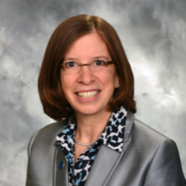

Dr. Maria Richman
Low Vision Rehabilitation Optometrist, Shore Family Eyecare

This Macular Chat episode featured Dr. Maria Richman, a Low Vision Rehabilitation Optometrist from Shore Family Eyecare in New Jersey. Dr. Richman discussed Advanced Macular Degeneration and provided an update on the investigational medical device called the SING Implantable Miniature Telescope. She discussed the improvements and benefits made to the new implantable telescope device as well as the criteria that is needed in order to join the clinical trial for the device.
Download English Transcript PDF
MS. DIANA CAMPBELL: Hello, and welcome to this month’s BrightFocus Chat about macular degeneration. My name is Diana Campbell, and I am pleased to be here today with this community of people who are impacted by macular degeneration. We will also be using the term “AMD” throughout the Chat, which stands for “age-related macular degeneration.” The topic of today’s Chat is “Advanced AMD: An Update on Investigational Medical Device.” We are going to spend about a half an hour learning about the potential benefits of the device and the clinical trial studying it and answering your questions. For those of you who are new to our Chat series, this Chat is brought to you today by BrightFocus Foundation. We fund some of the top scientists in the world who are working to find better treatments and, ultimately, cures for macular degeneration, glaucoma, and Alzheimer’s disease, and we do events like today’s Chat to get the latest news from science as quickly as possible to families that are impacted by these diseases. You can find much more information on our website, www.BrightFocus.org.
We are delighted to host today’s guest, Dr. Maria Richman, a low vison rehabilitation optometrist from Shore Family Eyecare in New Jersey. Along with treating her patients, Dr. Richman has presented research and lectured at many optometric, educational, and rehabilitation conferences. Dr. Richman, thanks so much for joining us today. Before we get into the discussion of the investigational device that’s being implanted through a mini telescope, can you tell us a bit about what a low vision rehabilitation optometrist does and the services you provide at your practice or for people with AMD?
DR. MARIA RICHMAN: Yes, Diana. Thank you. Well, hello, listeners. My name is Dr. Maria Richman, and I am a low vision doctor—a rehabilitation optometrist. Who is a low vision doctor, and what do they do? Well, a low vision optometrist is a type of eye doctor. We focus on improving the patient’s visual function, including the activities of daily living. So, what do I do during a day as a low vision doctor? I deal with patients by providing a comprehensive low vision exam to help identify the healthy and unhealthy areas of the eye, and I take measurements to prescribe optical and non-optical devices. Then, I provide training and therapy techniques to help patients maximize their remaining vision and function better, and I also help patients understand their limitations and I help them to establish realistic goals. So, when you go to your eye doctor, sometimes you turn to your eye doctor for medical intervention, but also know that you can turn to your eye doctor for visual and functional intervention as well, so that’s how eye doctors, in general, work. Some function or specialize more on medical eye care, and others specialize or pay more attention to visual eye care, and so that’s a little bit about what I do as a low vision eye doctor.
MS. DIANA CAMPBELL: I really appreciate that. It’s a question that we get a lot, and I think the experience of many people is that they don’t necessarily get a referral or don’t know how to find one, so the outline of what doctors like yourself do is really important. And I’m pretty sure we’ll get some questions at the end of the Chat, too. So, let’s go over the basics about the SING implantable miniature telescope—or the IMT as we might call it throughout the Chat. Consuela from Connecticut asks, “What is the benefit of the device?” So, could you tell us a little bit about the device, how it works, and your involvement with the CONCERTO clinical trial?
DR. MARIA RICHMAN: Yes. First, I’d like to give you a little bit of a history on the device. The IMT, which is the implanted miniature telescope, first was proceeded by a first-generation IMT back in 2010, so the first-generation IMT, which is the implantable telescope, has similar optics to the current telescope, which is in the study that we’re dealing with or discussing today. The first generation was implanted in more than 700 patients with macular degeneration, and over a 5-year period, we found that 90 percent of the patients with the implantable telescope achieved at least a two-line gain in their visual acuity.
We also looked at the patients with the first-generation implantable telescope device, and we measured results from the National Eye Institute questionnaire—that’s the NEI VFQ-25—which had questions about general vision, near activities, distance activities, social functioning, mental health, and more. And the mean scores from that questionnaire show increases in seven out of the eight relevant categories, with patients reporting on less dependency on others; being less limited in their everyday activities; and being able to recognize faces quicker, easier, and follow more details. So, when we saw how much good came out of the first-generation device, we said, “How could we make it better?” And in making it better, we improved the surgery and delivery of the device so that now we have a smaller incision that’s made with less sutures, which leads to a quicker recovery and people seeing better sooner. We’re finding post-operatively—1 to 3 days later—people are seeing better. And what that translates to is that they can be connected to the low vision doctor—the rehabilitation team—sooner, so that we can give them the tools they need to succeed by working with their low vision doctor.
So, that’s the history of how we got to where we are today. It’s by streamlining something that already works and making it work better. And by making it work better, we’re helping people to use new vision or new visual techniques to be able to see details and activities that, maybe, they’ve had to give up on because their sight … their center vision sight had been failing from late-stage macular degeneration. Does that answer your question, Diana?
MS. DIANA CAMPBELL: It did. I was just going to follow up with: How does the device actually work? So, you have the surgery. Could you outline which eye it goes into and how it helps you to see better?
DR. MARIA RICHMAN: Yes. Well, what happens with the process is that there’s many team players helping the patient to be successful. So, when we start the beginning of the patient’s journey, we are having the retinal doctor work with the patient, the cataract surgeon work with the patient, and the low vision doctor work with the patient. So, we want first … well, in any order, we want the patient to be cleared by the retinal doctor that they truly have the late-stage macular degeneration, which is the dry form—it’s the late-stage AMD. They need to have results from either the dry AMD or the inactive wet AMD, so that we’ll be able to get clearance from the retinal doctor.
So, once again, it is a dry form of AMD, and it’s a late-stage AMD that would be the criteria that needs to be met for the patient to be entered into the study. The type of AMD has to have the bilateral geographic atrophy or the disciform scar—the scarring that happens in the macular area. The patient also has to be cleared by the cataract surgeon, so they have to meet certain corneal health requirements. And, of course, they have to still have the lens in their eye. They cannot already have had cataract surgery because to put the implant into the eye, the cataract or the lens of the eye still has to be intact so that when the cataract surgeon comes to do the surgery to implant the telescope, first we have to remove the lens or the cataract from its capsule and then the implantable miniature telescope gets delivered through the surgical procedure into that area where the cataract has just been removed.
So, you see, we will need retinal clearance that the type of late-stage macular degeneration, the type that would meet the criteria for the study, and we also need to check with the cataract surgeon to make sure that the eye is healthy enough to remove the existing cataract and place the miniature telescope into that position in the eye. And then, of course, they need to visit their low vision doctor, which would be myself—if you are living in New Jersey, you would get to meet me—and what I do is I have what is called a telescopic simulator. I have an external telescope that you would hold in your hand, and it’s a similar magnification to the actual implanted telescope that we would be using during the surgery. But before we get that close to the surgery, we want to first see in the exam lane that you’re comfortable in seeing through a handheld telescope of the similar power so that you could appreciate that the magnification would give you enough detail to recognize faces, watch TV, look down the street, see someone waving at you from across the hallway or down the food aisle, or so forth.
So, we want to first make sure that you have an appreciation for the level of magnification that the telescope would be providing for you, as well as understanding what bi-ocular viewing is, because what happens in these cases with the implantable telescope, Diana, is that you would hold the external simulator—the external telescope—up to one eye, let’s say the operated eye, so that you could appreciate the enlarged, clear central vision that you get with that eye, and then maybe you’d wink or blink a little bit there so that you concentrate on the other eye—the unoperated eye—so that you could see around the room. So, one eye would be delivering you central, enlarged, clear, recognizable vision, and the other eye—the unoperated eye—would provide the visual field information, the side vision information, the mobility information of where you are in space and how comfortable you are in walking around with your regular vision that you’re used to in the unoperated eye.
So, it will take some training to make sure that you understand that it would be a new way of seeing; it would be a new type of viewing—the bi-ocular viewing—where one eye would be paying attention to central vision and the other eye would be paying attention to peripheral vision. And then, once you’re clear with the low vision doctor, the cataract doctor, and the retinal doctor, then you’re ready to have your surgery. And so, the surgery would just be a surgical procedure that the cataract surgeon would remove the cataract, place the implant into the eye. It requires just a few sutures, and then you’d have follow-up post-op care with your surgeon, and in a day or two, you’re seeing better, and by 2 weeks after the surgery, you’re in your low vision optometrist’s office, or you’re rolling up your sleeves with me and we’re going to do a visual acuity assessment. I’m going to provide optometric care through that comprehensive eye exam that I spoke about earlier when I was introducing myself, and then we’ll start the vision rehabilitation management, which can take about six to eight sessions of rehabilitation or vision therapy or eye exercises.
And the roles … that would be in-office eye exercises as well as home exercises. So, over the six to eight sessions of working closely with you, your low vision specialist would be helping you to function better with your new vision, helping you to use your vision more easily and more comfortably. We’ll be teaching you new visual tasks and techniques to help you to see better and motivating you throughout this whole process, because it’s like anything else. It takes work and it takes motivation, and it takes concentration, and it also takes a good support network. So, we do ask for patients who are interested in going this route with us that you have a good support network in place, either with caregivers or families or friends or other people in your life because we want your support network in the office with us so that they can see the techniques we’re doing to train you and give you tips on how to maximize your new vision, and for someone who can help you with your home exercises. Because not only in the office are we going to do therapy, but you’re going to do therapy at home as well, so it’s almost as if you’ve had a hip replacement or a knee replacement. After that replacement, you just don’t start walking or running, you know, you have to go for PT—for physical therapy. You have to start working that leg, working that knee, working that hip, and learning how to walk again with a device, a prosthetic, or something new in your body. Well, it’s going to be the same thing when we put the implantable telescope in. It’s something new in your body that just takes some work and encouragement and motivation for you to stay in the process and do your exercises and keep moving along.
MS. DIANA CAMPBELL: That’s great. That’s a great analogy with a knee replacement or a hip replacement. You definitely highlighted how important it is to have that rehabilitation in place and to stick to it and do the exercises to get the most use of the vision that you have. I know we talked a little bit about managing expectations, and it sounds like it’s really seeing the people that you love and being able to do some hobbies again and that sort of thing that people will be able to regain. I just wanted to mention expectations here and have you tell us: Are we expecting 20/20 vision or driving, or is it really just those activities and things that people love to do and are no longer able to?
DR. MARIA RICHMAN: Correct. Well, that’s a great lead in, Diana, because I wanted to touch on that a little and I said, “Let’s wait until we have enough time to pay attention to that.” So, if you remember, when I introduced myself as a low vision doctor and what I do, not only do I find the healthy and unhealthy areas of the eye and then I take measurements to give you the tools to succeed with either prescribing optical or non-optical devices, but I do the therapy and the training with you to help to teach you how to maximize your remaining vision.
But what else I do is I help you to establish realistic goals so that we’re both on the same page, because we’re a team at this point. We’re both going to roll up our sleeves and we’re both going to work to get you to the goals that we agree are realistic. So, what are the realistic goals for a patient that’s interested in the implantable telescope—the SING IMT? The IMT … the SING IMT is the small-incision, new-generation implantable miniature telescope. If you come to me and say, “Doc, I heard your BrightFocus Chat. I love it. I went through the screening. They told me I could come to you. I want to drive.” And I’m going to say, “That is not a realistic goal of what the implantable telescope is for.” The implantable telescope is to bring people more connected visually to their everyday activities. So, realistic goals would be watching TV again, recognizing people, seeing your grandchildren’s faces, sorting through … somebody just got married and sent you a bunch of pictures because maybe you couldn’t have gotten to the wedding, but you want to see pictures of your granddaughter; so, seeing pictures, going out to social events again.
You know, so many times macular degeneration robs you of being comfortable out at social events because so many people from across the room might be waving to you but you can’t see them or you can’t recognize them, and you don’t want them to feel like you’re not paying attention to them or you’re ignoring them because the center vision from macular degeneration robs you of being able to recognize faces, so little by little people give up going to social events because they feel awkward around people. The SING IMT, the implantable telescope, gives people back the ability to recognize faces, to see people, to get out and socialize again. It also helps you to bring back your hobbies, so the things that you used to do during the day that made you feel more complete and more purposeful. We can work on hobbies again. We can help you with reading and writing and near activities and getting around in social settings. So, those are all realistic goals. But I’ll tell you, Diana, if people came to me and said, “Doc, I want this telescope because it’s going to give me clear vision immediately.” I’d have to say to them, “I’m sorry, it does give you clear vision, but it takes a lot of work.” It’s almost like that knee replacement. “Doc, I want a new knee so I can go out dancing or running again, but I don’t want to do … I don’t have any time for PT—for physical therapy.” Well, that doc’s going to tell you the same thing. This is not for you if you’re looking for some clear, immediate gratification. It’s going to take work. It’s a process. It’s a pleasurable process, but with work, you know, you have to have the commitment. You know, some people might say to me, “Doc, I want to get back to playing tennis or seeing a golf ball in flight so I can see where it’s heading and where it’s landing.” This type of implantable telescope is not for that. It is more … we want to take baby steps. We want to regain our new vision little by little and see where that takes us.
So, I would redirect those patients that might have unrealistic goals of clear, immediate vision or driving vision or seeing a golf ball in flight. I would redirect those goals and see if we could come to some common agreement that … let’s start by watching television again, getting out and socializing again, even going out to lunch with your friends and being able to read a menu or cut the food and not feel like you can’t see enough and you might make a mess at the table, you know? There are little things that we’ve just given up and accepted not having them in our lives anymore because our vision is failing, and now we can bring that independence, that confidence, back by socializing, being around people, catching up on TV programs, spending that quality time of knitting or doing model painting or putting puzzles together or just sorting through your mail. You know, reclaiming that this is my piece of life that I want to be able to control again instead of always having to wait for someone to come to my house and sort through my mail and tell me what’s in the pile that I’ve accumulated over the week. Now, you have more confidence to feel like what is a part of my life is what I can control? And maybe I’ll still need some help from family and friends and maybe I still will need a magnifier but maybe it won’t be as strong or maybe it won’t … maybe I won’t always need to use that magnifier in different settings, but I have that confidence that I can get through the activities that are important to me every day and I can do that for the most part by myself. Those would be the realistic goals that an implantable telescope could give a patient again.
MS. DIANA CAMPBELL: Thank you for that. I think it’s so important for people to know what they’re getting themselves into and what they can expect, but also learning how they can get some of that independence back. I know that’s one of the biggest frustrations and it leads to anxiety and depression and all kinds of other things—it’s just that loss of independence, which is so important to us all. I’m getting so many questions. So, my next question … the next thing we’ll discuss will be the study criteria for the clinical trial—the participant criteria—who’s eligible, who’s not eligible. Some of the questions that I’ve been receiving that we should cover are: Why does previous cataract surgery disqualify you from the implant and whether that cataract surgery is in the eye that would been to the operated or not, and then a bunch of questions about wet AMD and injections. So, if we could talk about the criteria and cover those as well.
DR. MARIA RICHMAN: Great. Okay, well, I jotted down the notes because it had about three different sections in my brain of what I had to touch on, so let’s start with the criteria first. Just so we go back to the very beginning of this discussion, this is an update on an investigational medical device. So, this is an FDA clinical trial, and I am one of the investigators, and with the FDA clinical trials, it is very rigid, it is very specific, it is very reproducible because we have to stay in the parameters of what the criteria are. So, if they list X, Y, and Z as the criteria and you say, “Well I have X and Y but not Z, can I still be in it?” And it’s “No, it’s an FDA clinical trial.” We have to go step by step specifically on what the criteria pieces are. So, I’m going to go through the criteria of who is eligible for the IMT, the implantable telescope. We have an age criteria, so it has to be someone 65 or older. That person has to have the late-stage AMD resulting from either dry or wet AMD, but the wet AMD has to be inactive for at least 6 months, and we also need … and that’s what makes it the late-stage AMD. It’s either that it’s dry or the inactive wet, where it has not been wet for the last 6 months, and there has to be a presence of this bilateral geographic atrophy, or the bilateral scars, where there’s a blind spot or there’s a missing spot, like when you look at somebody’s face, like a piece of their … you know, like their face, maybe around one eye, is missing or the part where their mouth should be is missing. When you have those blind spots from macular degeneration, that’s that disciform scar or that geographic atrophy—that bilateral geographic atrophy—that we’re talking about.
Now, I know previous cataract surgery that was part of that second question, the part of this study is that the patient has not had cataract surgery in the potential IMT implant eye. So, let’s say we determine that your right eye would be the implant eye that you would benefit the best from having the implant placed in—having the miniature implantable telescope placed in your right eye. That right eye cannot have already had cataract surgery. That’s just the requirement of this FDA study, so you would have to be a patient who has a cataract … and I’m just picking the right eye. That doesn’t mean that every case everybody has a right eye that has the implant in, I’m just making an example here. But to make things clear, the implanted eye must not have had cataract surgery—previous cataract surgery—in that eye for that eye to be a potential IMT implant eye—the implantable telescope eye. Also, another criteria is visual acuity. So, with the visual acuity, the criteria have to be around 20/200 or worse, and I think most of you understand that 20/200 level because 20/200 or worse—that is according to the Federal Disability Act—that is the definition of legal blindness, right? So, we’re looking at specific criteria. If you wanted to write it down, or at least it will be in the recording, so when you go back to listen to this, the specific visual acuity criteria are 2/160 to 2/800 in each eye. But all you need to know when you go to talk to your doctor is, “Doc, am I in the ballpark of 2/200 or worse?” If that’s the case, then that would be one of the criteria we’re looking for. I also just want to go back to the cataract surgery question because I don’t know if I made myself clear. We cannot … for this study, we cannot remove the artificial lens—the artificial lens in the eye—that was placed in the eye from the cataract—from a previous cataract surgery. So, if you’ve already had cataract surgery and you had an artificial lens placed in—which is that IOL, interocular lens placed in already—for this study, we cannot remove that and place in the telescope. So, I just wanted to go back to the cataract surgery question earlier.
MS. DIANA CAMPBELL: You answered the question before I could ask it.
DR. MARIA RICHMAN: Okay. I read your mind; you read my mind. Good. Okay. The other criteria … the other part to the criteria would be we need adequate peripheral vision in the non-operated eye. So, the eye that does not get the telescope implant in it, we want to make sure that that fellow eye—that other eye—has good peripheral vision because now that’s going to be the workhorse for gathering peripheral vision because the telescopic eye is going to be the workhorse for gathering enlarged, central, detailed vision. And then, we also want to make sure that the cornea is healthy enough to meet the requirements, and we also want to make sure that the retina is healthy enough to meet the requirements, because we already know that the retina can have the late-stage macular degeneration; that’s part of the requirements, so that’s okay—the late-stage macular degeneration. But if you have other retinal conditions in addition to your macular degeneration, conditions, like diabetic retinopathy or a history of retinal detachments, or other retinal conditions, like Stargardt disease or retinitis pigmentosa—if you have a history of other retinal or retinal vascular diseases in addition to your macular degeneration—then you would not be a candidate. You would not meet the criteria because part of the criteria is that you just have the late-stage macular degeneration. So those are the key points. There’s a lot more to the inclusion criteria of who is eligible and who are not eligible, but still to the key points: the age; the presence of the late-stage macular degeneration, the presence that there still is a cataract, that the eye has not already gone through cataract surgery; that the visual acuity is around 2/200 or worse; and that the fellow eye—the non-operated eye—has adequate peripheral vision—the eye that’s not receiving the implant has still good side vision because, as I said with that bi-ocular viewing, you need one eye for central vision, which would be the telescopic eye, and then the other eye is the non-implanted eye and would be the eye for peripheral vision. Peripheral vision, that helps you with mobility purposes so you’re safe walking around, you don’t bump into things, and that you know where you are in space, you know your orientation, you know there’s a chair in the corner, you know that the door is over to the left side of the wall, things like that. So, if you need—
MS. DIANA CAMPBELL: I also want to clarify when you said no active wet AMD. I believe—and correct me if I’m wrong or let’s share this with everybody—essentially that means no injections in the wet AMD eye for, I think, it was 6 months before the surgery?
DR. MARIA RICHMAN: Correct. Correct Very good point, Diana. Yes. So, maybe let’s get back. Let’s put the FDA study and the implantable telescope just to the side for now. Let’s talk about age-related macular degeneration, also known as AMD, I keep on referring to. AMD damages the macula. The macula is the area of central vision in our retina; it’s our detailed vision area. It’s our area of 20/20 vision. When that is damaged, we can’t see faces anymore, we can’t recognize people as easily anymore, it’s difficult to watch TV, it’s hard to read your mail, you can’t always set your thermostat, you can’t always read a recipe, it’s hard to read the label on your medicine bottles, it’s hard to go out shopping to see a price tag or to read a menu if you’re going out to eat, all right? All of these problems stem from damage to the macula. And in macular degeneration, AMD—the age-related macular degeneration—AMD starts as a dry form; it starts as dry AMD. In some cases, the dry AMD advances to a more severe dry AMD; it takes up a larger area of the macula, but the area is a dry condition. In other cases, the dry AMD changes to wet AMD, and when you have wet AMD, that may require injections. Now, for this study, the AMD must be dry or an inactive wet AMD. Now, an inactive wet AMD is one that does not need any more injections. It’s changed back to a dry AMD, that’s why you’re not getting injections anymore, and it’s at that point when the AMD has become this late-stage AMD that, at that point, you meet the criteria for this FDA study. Does that explain it a little bit better, Diana?
MS. DIANA CAMPBELL: It really does. Thank you so much for that clarification. So, let’s say someone is eligible with all the criteria and elements we just discussed, what would one expect from the surgery itself? Is it inpatient? Outpatient? You kind of mentioned or touched on the recovery time, but what is the process itself? How long does it take, and where would they do this?
DR. MARIA RICHMAN: Okay. In New Jersey, we have two locations—we have two surgeons in New Jersey—and I’ll go over all different sites that we have so far. But with the surgical procedure, it includes the cataract removal with the insertion of the implantable telescope. It does require an incision so that the delivery—the preloaded delivery system—can place the telescope properly in the eye. It does require a couple of sutures and postdoc care. Usually in a couple of days you’re seeing better, by 2 weeks you’re usually free from the surgeon, and you’re then in the low vision doctor’s office so that we can start the rehab together. The surgery is similar to cataract surgery, so you would go to the cataract surgeon’s surgery center. It takes a little bit longer than a regular cataract surgery does, but it’s basically the same steps. It’s just a little extra time because it’s a delivery system which is different than the interocular lens, but it’s very similar; it just takes a little bit more time. It takes a little bit more sutures than the standard cataract surgery, what the implant does, but by 2 weeks you’re free from the surgery and the surgeon and you’re in the low vision optometrist’s office doing the rehab.
MS. DIANA CAMPBELL: Just to make it perfectly clear, they’re not going to be in the hospital or something like that for 2 weeks.
DR. MARIA RICHMAN: No.
MS. DIANA CAMPBELL: That’ll just be the expected recovery time for the procedure?
DR. MARIA RICHMAN: Correct. They’ll have the surgery in the doctor’s surgery center, and they go home that day, and the next day … and the next day or couple of days they’re already seeing better, and by even maybe sooner than 2 weeks, but we usually say 2 weeks. It’s 10 days, 2 weeks; by that time, you’re done with the surgeon. So, yes, you don’t go to the hospital, just like you don’t go to the hospital for a regular cataract surgery; you usually will just go to the surgery center that the cataract surgeon has. So, all that is similar, so if you’ve already had cataract surgery maybe in one eye—the eye that is not being evaluated for the implant—then you already know the steps to the cataract surgery procedure, so it will be similar. And as I said, that’s the key. That’s why this is called SING, the SING IMT. It’s smaller incision, new generation—that’s how we made it better from the first generation is by improving the surgery. So, the incision site is much smaller, the number of sutures is much smaller. They’re saying that the procedure takes maybe around 30—30, maybe 40—minutes; and post-op care, usually 1 to 3 days, the patient’s already seeing better. So, post-op care is very quick, and recovery, especially in seeing better, is rather quick as well. So, that’s the exciting part of why this is called SING IMT, because it’s … the new generation is we’re using a smaller incision, less sutures, quicker recovery time, seeing better sooner, and getting you to the rehab programs sooner.
MS. DIANA CAMPBELL: Wonderful. We’ve got time for about two topics that we’d like to cover, and one comes as a question from Kay from California, “When will my retina specialist have access to this or where can I find out if I can be part of the trial or receive the device?” And then, I think we can walk through again how they can find out about the trial and the sites and conclude with that.
DR. MARIA RICHMAN: Great. No one really has asked so far, “How is this working so far?” Well, we have been … this has already been done worldwide, and we have over 100 patients with the SING IMT—the small-incision, new generation implantable telescope—to date, so we have over 100 patients already worldwide that are reaping the benefits from the device and the rehabilitation that goes hand in hand with the device, so that’s exciting news. The way we are with this FDA study is that we have locations across the United States. In New Jersey, we have three locations; in California, we have two locations; we also have locations in Massachusetts and in Florida, in North Carolina, in Wisconsin, and in Texas. We are in conversation with the retinal community, with the cataract community, with the low vision community, so mostly all the areas where macular degeneration patients will be going to continue their everyday care. Their doctors will be reading about this FDA study, will be learning about this FDA study, will be following the progress of this FDA study. We are reaching out to regional groups and national organizations so that we’re reaching as many doctors and health care providers who are involved in the care of macular degeneration patients.
Your retinal doctor probably is already aware of this, but we also will go over anyone interested to learn more about this study and about if they would be a candidate for this implantable telescope. We have a phone number and a website; we’ll go over that at the end of the discussion. Once you either call the phone number or visit the website that we give you, there will be a member from our team or a connect team that will do a phone screening with you to see if you qualify for the study. And, again, remember the criteria for the study would be someone 65 or older who still has a cataract in one eye and that they have the late-stage AMD that results from either the dry AMD or the inactive wet AMD. And then from there, we would … you would get put through the process of being educated if you meet the criteria from being evaluated by the low vision doctor, by the retinal doctor, by the cataract and corneal doctor because, of course, like any FDA study or any interaction that a doctor has with a patient, we only want to involve the people who will be a success.
So, we never want to put you in a situation where there is something that wouldn’t help you to reap the benefits of this device, and that’s why the criteria are so strict is because we … fortunately, we already had the first generation, which was successful—we’re just making it more successful. So, that’s a good thing, that we have so many good things that already came out of the first generation by changing or tweaking the surgery a bit. That’s helping everybody to have less sutures, less healing time or a quicker healing time, and quicker time to get to the vision rehab doctor—the low vision specialist—so that you can learn how to use your new vision quicker, easier. And the study goes on for a little over a year, so it is a time commitment, and that’s why we want people who are interested in this study to know that you have to have the motivation and the support team because we are looking at a 12- to 15-month study length of this FDA clinical study. So, it’s a time commitment, but as you can tell from the areas that I highlighted, if we can improve your visual function, if we can help you to use your new vision better, if we can give you therapy techniques that can help you to master seeing better and clearer and more efficiently and keep you motivated and driven through this whole process with caring doctors and caring support staff and a caring network that you bring to the experience as well with your caregivers and with your families and your friends that’s all … it’s a process; it’s not a quick fix.
So, please know that we’re interested in you contacting our team to discuss eligibility and potential participation and to answer any of the questions that you have that maybe weren’t answered during this time during the Chat, and again, we have five visits in approximately 12 to 15 months, plus the vision rehabilitation that you go through. For those who have a pen and paper, I’ll give you the phone number over the phone now, but we’ll also be able to get it to you again through other means. But for those who want to write down the number of how to contact us to talk to our team members, our number here is 1-866-393-3767. Again, to contact us and talk to your team members to discuss eligibility and participation, or if you just need some questions answered a little bit more in depth than what I was able to do today, call us at 1-866-393-3767. And we also have a link, which is www.ConcertoStudy.com, and Concerto Study is C-O-N-C-E-R-T-O-S-T-U-D-Y. And as I said, we’ll get this all to you in case you can’t write this down now. You’ll still be able to connect with us after the Chat is done today.
MS. DIANA CAMPBELL: Thank you so much. This is so much information, and I know we’ll get lots of questions on the voicemail afterward, and we’ll follow up with folks. We will provide the phone number and the website again, and I’m just really excited about this. A couple of final notes before we conclude. Next month we’ll be taking a break for the holidays, and we’ll be back after the new year on Wednesday, January 25 at 1 p.m. Eastern. We’re actually currently planning our list of topics to cover in 2023, and we’d love to hear your suggestions or ideas, so you can also leave those on the voicemail after the call. We also have our next Glaucoma Chat in the new year on January 11. If you have already registered for the Glaucoma Chats, you’ll just automatically receive that reminder call the day before the Chat. If you’re interested in listening and you haven’t already registered, you can also leave a message after today’s Chat. So, to close out today, Dr. Richman, are there any final remarks you’d like to share with the audience?
DR. MARIA RICHMAN: Well, I always want to remind low vision patients and people in general, if you have macular degeneration, glaucoma, diabetes, whatever you have that has weakened your eyesight, just know that to never to give up hope. Your doctors have not given up hope and neither should you, so always know to ask questions, to have the conversations with your doctors, to be educated by listening to BrightFocus Chats like we’re doing today so that you understand that there’s lots of research out there, that we know that people are out there suffering, that we feel your pain. We know especially with macular degeneration there’s more than 11 million people who are affected by some form of macular degeneration in the United States. We know there’s a problem. We’re trying to fix the problem. We cannot cure the problem, but we are working very hard to bring more functionable, usable vision back to your life so you can have a better quality of life. So, please don’t give up on yourself; we are not giving up on you. Always have hope, always have questions, always try to educate yourself, and always know that the medical community is working very hard to help you. Thank you, Diana.
MS. DIANA CAMPBELL: Thank you so much. This has just been so wonderful, and I think those final remarks are hopeful and remind people that, while it might be slow-going, everyone is working really hard in the research and medical community to really find some better solutions. So, with that, this will conclude the BrightFocus Chat. Have a wonderful holiday season, and we will be back in January. Thank you.
DR. MARIA RICHMAN: Thank you, Diana.


BrightFocus Foundation is a premier global nonprofit funder of research to defeat Alzheimer’s, macular degeneration, and glaucoma. Since its inception more than 50 years ago, BrightFocus and its flagship research programs—Alzheimer’s Disease Research, Macular Degeneration Research, and National Glaucoma Research—has awarded more than $300 million in research grants to scientists around the world, catalyzing thousands of scientific breakthroughs, life-enhancing treatments, and diagnostic tools. We also share the latest research findings, expert information, and resources to empower the millions impacted by these devastating diseases. Learn more at brightfocus.org.
Disclaimer: The information provided here is a public service of BrightFocus Foundation and is not intended to constitute medical advice. Please consult your physician for personalized medical, dietary, and/or exercise advice. Any medications or supplements should only be taken under medical supervision. BrightFocus Foundation does not endorse any medical products or therapies.

Gene therapy treatments may one day free people with age-related macular degeneration (AMD) from the need for frequent eye injections.

In recognition of National Caregivers Month, this episode explores the vital role of those who support individuals living with vision loss—whether family members, professionals, or volunteers.
Dr. Jeffrey Stern and Dr. Sally Temple, Principal Investigators and Co-Founders of the Neural Stem Cell Institute, will explain what stem cells are and share the latest updates from clinical trials.
Artificial vision systems utilizing these technologies show promise for individuals with profound vision loss in early trials.
Join Dr. Sara Fard, a retina specialist at Illinois Retina Associates, as she explains the benefits of sustained GA treatment, including slowing the rate of vision loss, protecting retinal tissue, and supporting daily visual function.
Help Fight Macular Degeneration and Save Sight
Your donation helps fund critical research to bring us closer to a cure for this sight-stealing disease and provide vital information to the public.
Donate Today