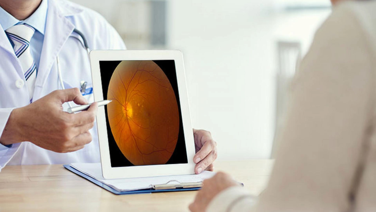
Learn why the distinction between hard and soft drusen is important, and how the type of drusen that you have may influence your risk of vision loss.
People are diagnosed with age-related macular degeneration (AMD) when they have drusen detected in their eyes. The ophthalmologist may call these “hard” or “soft” drusen.
What are Drusen?
Drusen are tiny deposits under the retina that the eye doctor can see during an exam. They can also be photographed with a retina camera or using an imaging device called optical coherence tomography (OCT). This latter image shows a cross-section of the retina and underlying drusen.
Drusen are about the width of a pinhead, and are composed of a mixture of proteins and lipids (naturally occurring molecules that include fats). They often cause no symptoms, but can occasionally cause visual distortion if they are very large and near the center of the retina.
Size and Number of Drusen
The size and numbers of drusen are very important. Larger numbers of drusen, as well as drusen of larger size, indicate higher risk for some vision loss in the future.
Hard vs. Soft Drusen
“Hard” drusen are small, and indicate lower risk of future vision loss than “soft” drusen, which are larger, cluster together, and have edges that are not as clearly defined. Whereas the chance of a person with a few hard drusen losing some central vision from AMD in a 5-year timeframe is only a few percent, the chance may be as high as 50 percent over the same time period for those with many intermediate and large-size soft drusen.
Prevention of Vision Loss
One can lose some central vision if their AMD progresses to one of the advanced forms: wet AMD or geographic atrophy. Since the risk of developing advanced AMD is elevated in people with many intermediate and large soft drusen, it is recommended that these individuals take AREDS2 formula vitamins, which decrease the risk of progression to wet AMD by about 25 percent.
Those with drusen, especially if they are soft drusen, should monitor their vision at home, checking their vision on an Amsler grid, which can detect new onset wet AMD. This should be done one eye at a time, with the other eye closed. If wet AMD is detected early, there’s a good chance that vision can be stabilized or even improved by injection of the medications brolucizumab (Beovu®), aflibercept (Eylea®), ranibizumab (Lucentis®), or Avastin® (bevacizumab).
People with any type of drusen can decrease their risk of AMD progression by avoiding smoking, minimizing red meat and snack foods, and eating a diet high in vegetables, fruits, and fatty fish (salmon, sardines, mackerel, or tuna).
Research Findings
Researchers have tested methods for eliminating drusen. One technique, using a laser, did not decrease the risk of vision loss. Another small study enrolling people with very large drusen near the center of the retina showed that relatively high doses of a statin drug were associated with drusen shrinkage and vision improvement. Further studies are needed before this approach can be widely recommended. Additional research into the causes of drusen and the reasons for their connection to AMD progression should lead to new protective approaches.
About BrightFocus Foundation
BrightFocus Foundation is a premier global nonprofit funder of research to defeat Alzheimer’s, macular degeneration, and glaucoma. Through its flagship research programs — Alzheimer’s Disease Research, Macular Degeneration Research, and National Glaucoma Research— the Foundation has awarded nearly $300 million in groundbreaking research funding over the past 51 years and shares the latest research findings, expert information, and resources to empower the millions impacted by these devastating diseases. Learn more at brightfocus.org.
Disclaimer: The information provided here is a public service of BrightFocus Foundation and is not intended to constitute medical advice. Please consult your physician for personalized medical, dietary, and/or exercise advice. Any medications or supplements should only be taken under medical supervision. BrightFocus Foundation does not endorse any medical products or therapies.
- Disease Biology
- Prevention


