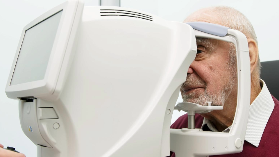
The past decade has witnessed a revolution in retinal imaging providing detailed views of retinal changes in people who have age-related macular degeneration (AMD). Learn about the various forms of retinal imaging and how they help eye doctors diagnose and treat AMD.
New retinal imaging techniques have been driven by advances in optics, cameras, computers, and software, and have become extremely helpful guides for the diagnosis and treatment of AMD.
Retinal images have become so valuable that people with AMD can expect to have at least one, and sometimes several, types of images taken on each visit to the eye doctor.
Retinal imaging techniques used to diagnose and treat AMD may include:
- Optical Coherence Tomography
- OCT Angiography
- Retinal Fundus Photography
- Autofluorescence Imaging
- Scanning Laser Ophthalmoscopy
Each technique is discussed in more detail below.
Optical Coherence Tomography
The greatest advance in retinal imaging has come from optical coherence tomography (OCT). This technique uses infrared light reflections off the retina to generate a cross-section retinal view that is displayed on a computer screen.
The images are acquired within a few seconds and do not require any uncomfortable bright light flashes. They show pockets of fluid in the retina in patients with wet AMD, helping to guide the frequency of anti-VEGF injections (Beovu®. Eylea®, Lucentis® or Avastin®). They also show drusen, which are small deposits under the retina in patients with early dry AMD, as well as retinal thinning from cell death in patients with late dry AMD (also called geographic atrophy). Learn more about a leading scientist funded by the BrightFocus National Glaucoma Research program who helped develop OCT.
OCT Angiography
A recent improvement to OCT technology is OCT angiography. With this technique, normal and abnormal retinal blood vessels can be seen. This helps the ophthalmologist understand the nature and extent of abnormal blood vessels in people with AMD.
Until OCT angiography was invented, detailed views of retinal blood vessels could only be obtained following intravenous injection of dyes, called fluorescein or indocyanine green (ICG). These tests are more uncomfortable, as they require the injections and sometimes light flashes over a period of 15-30 minutes. However, the tests are still required at times to obtain an accurate and complete picture of the disease process.
Retinal Fundus Photography
Retinal fundus photography provides a color picture of the retina. This technique has existed for decades, but has been improved by digital cameras, computers, software, and new optical techniques. It takes only about a minute and requires a few bright flashes. The images obtained can show drusen, blood, or lipids (a group of naturally occurring molecules that include fats) that have leaked out of abnormal blood vessels in wet AMD, scar tissue, and areas where retinal cells have wasted away and died, called atrophy.
Autofluorescence Imaging
Atrophy can also be detected using autofluorescence imaging. This technique, which is also quick and requires just a few flashes of light, yields a view of the retinal pigment epithelial cells (RPE), which die in people who have geographic atrophy. RPE cells protect and nourish the retina, remove waste products, and provide a number of other functions.
Live RPE cells appear bright because they contain the autofluorescent material called lipofuscin, but areas in which the RPE cells have died appear as a dark patch in the photograph, since these areas no longer have lipofuscin.
Scanning Laser Ophthalmoscopy
An even sharper image of the autofluorescent RPE cells can be obtained with a scanning laser ophthalmoscope (SLO), but this imaging takes a bit more time and additional equipment. Even better resolution can be obtained with adaptive optics-SLO (AO-SLO), which borrows technology from high resolution telescopes. This literally uses a space-age technology that provides views of individual cells in the retina, allowing them to be counted. AO-SLO is current available only in research settings.
A relatively new optical advance within a camera made by Optos provides a view of the peripheral retina, rather than just the central retina (macula), and can be helpful in patients with suspected retinal detachment or other peripheral retinal abnormalities.
Talk with your doctor about what, if any, imaging techniques may be right for you.
About BrightFocus Foundation
BrightFocus Foundation is a premier global nonprofit funder of research to defeat Alzheimer’s, macular degeneration, and glaucoma. Through its flagship research programs — Alzheimer’s Disease Research, Macular Degeneration Research, and National Glaucoma Research— the Foundation has awarded nearly $300 million in groundbreaking research funding over the past 51 years and shares the latest research findings, expert information, and resources to empower the millions impacted by these devastating diseases. Learn more at brightfocus.org.
Disclaimer: The information provided here is a public service of BrightFocus Foundation and is not intended to constitute medical advice. Please consult your physician for personalized medical, dietary, and/or exercise advice. Any medications or supplements should only be taken under medical supervision. BrightFocus Foundation does not endorse any medical products or therapies.
- Eye Tests
- Treatments


