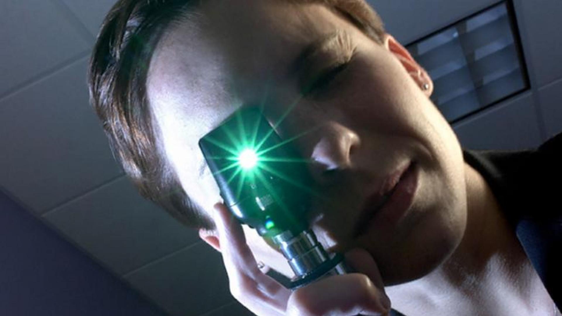
A macular hole is basically what it sounds like; a hole in the central part of the retina, which is called the macula. This hole causes blurring or distortion of the central vision. Learn about the similarities and differences between macular hole and macular degeneration.
What Causes a Macular Hole?
Macular holes can be full or partial thickness and can vary in size. They are caused by age-dependent contraction of the jelly in the center of the eye, called the vitreous. The vitreous is the substance that fills in the region from the lens to the back of the eye. As the vitreous shrinks, it pulls on the retina. In most people, the vitreous separates cleanly from the retina, but in some patients, it is tightly stuck to the retina and tears a hole. Then, fibers on the surface of the retina can pull on the hole to enlarge it.
Macular Hole Video
Macular Degeneration vs. Macular Hole
Macular hole occurs when some or all of the neurons in the center of the macula are pulled out of position. Macular holes are different from age-related macular degeneration (AMD), a disease in which deposits called drusen form under the retina and the light-sensitive cells (photoreceptors) of the macula slowly break down (dry AMD), or when abnormal, leaky blood vessels grow behind the macula and vision loss occurs from the death of the photoreceptors (wet AMD).
Treatment
Some small, partial thickness holes close without treatment. Others require outpatient surgery to prevent further vision loss, and, in some cases, improve vision. The surgery involves removing the vitreous jelly to stop it from pulling on the retina, and then replacing it with gas that holds the retinal in place as long as the patient is in a face-down position. This positioning may be necessary for about two weeks, until the gas bubble is absorbed and replaced by fluids made by the eye. Special devices are available to help patients maintain this face-down position. Following the surgery, clouding of the lens called cataract is common, and may require a second surgery to remove the lens and replace it with a clear plastic one. This, of course, is not an issue in patients who have already had cataract surgery.
Important Advice
Blurry or distorted central vision can be caused by a number of diseases, and patients who notice this should have a retinal exam by an eye doctor who uses dilating eye drops. Many people in their 40s and older (and sometimes in younger, very near-sighted people) will at some point see arcs of flashing light in their peripheral vision and floating spots like bugs flying across their vision when they move their eyes. These are symptoms of a vitreous detachment. The flashing lights only last a short time, but the floating spots, or “floaters” do not go away. They are most noticeable in bright light when looking at a white background or the sky. For most people this will not be a problem, but in a few, it may cause a macular hole if it tears the central retina, or a potentially a retinal detachment if it tears either the peripheral or central retina.
Patients experiencing sudden arcs of flashing lights at the edge of their vision, floaters, or a “curtain” blocking part of their vision should seek immediate attention from an eye doctor, as this can signal a retinal detachment, an emergency situation that can lead to permanent vision loss.
Treatment Overview Video
About BrightFocus Foundation
BrightFocus Foundation is a premier global nonprofit funder of research to defeat Alzheimer’s, macular degeneration, and glaucoma. Through its flagship research programs — Alzheimer’s Disease Research, Macular Degeneration Research, and National Glaucoma Research— the Foundation has awarded nearly $300 million in groundbreaking research funding over the past 51 years and shares the latest research findings, expert information, and resources to empower the millions impacted by these devastating diseases. Learn more at brightfocus.org.
Disclaimer: The information provided here is a public service of BrightFocus Foundation and is not intended to constitute medical advice. Please consult your physician for personalized medical, dietary, and/or exercise advice. Any medications or supplements should only be taken under medical supervision. BrightFocus Foundation does not endorse any medical products or therapies.
- Disease Biology
- Treatments


