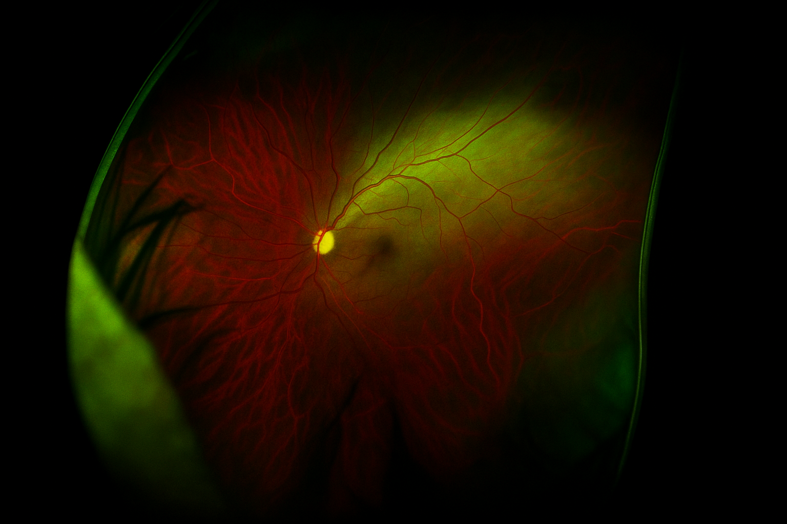
Retinal Ganglion Cell Axon Degeneration in a 3D Microfluidic Hydrogel Model

Principal Investigator
Shruti Patil, PhD
Indiana University School of Medicine
Indianapolis, IN, USA
About the Research Project
Program
Award Type
Postdoctoral Fellowship
Award Amount
$150,000
Active Dates
July 01, 2024 - June 30, 2026
Grant ID
G2024002F
Goals
This project aims to develop an advanced in vitro model using a 3D hydrogel-based biomechanical modulatory system on a microfluidic platform to assess increasing environmental stiffness on retinal ganglion cell axons.
Summary
Retinal ganglion cell (RGC) axons traversing the optic nerve head are highly susceptible to glaucomatous damage, yet the link between optic nerve head biomechanics and RGC axonal degeneration remains poorly understood. To address this, researchers will implement 3D platforms integrating microfluidics and hydrogels with tunable stiffness to model biomechanical aspects affecting RGC axons in the optic nerve head.
Unique and Innovative
The link between optic nerve head biomechanics and RGC axonal degeneration is poorly understood, necessitating an effective model to study tissue stiffness and RGC degeneration in a human cellular context. This proposal’s innovation lies in developing an in vitro human stem cell-derived culture system using a microfluidics platform with tunable hydrogels. This setup uniquely simulates biomechanical and structural attributes of the lamina cribrosa, previously absent in stem cell models of glaucomatous RGC degeneration.
Foreseeable Benefits
Upon completion, the study will uncover insights into RGC axonal dysfunction due to biomechanical changes in context of human tissue using a stem cell model. By targeting the biomechanical aspects of the lamina cribrosa, the study could lead to novel therapeutic interventions beyond traditional approaches regulating intraocular pressure. Incorporating hydrogels in the microfluidic system allows study of complex cellular interactions in a 3D microenvironment, applicable to other age-associated optic nerve degeneration. This offers hope for more effective glaucoma treatments.
Related Grants
National Glaucoma Research
Enhancing Access to Glaucoma Care Using Artificial Intelligence
Active Dates
July 01, 2025 - June 30, 2027

Principal Investigator
Benjamin Xu, MD, PhD
Current Organization
University of Southern California
Enhancing Access to Glaucoma Care Using Artificial Intelligence
Active Dates
July 01, 2025 - June 30, 2027

Principal Investigator
Benjamin Xu, MD, PhD
Current Organization
University of Southern California
National Glaucoma Research
Saving Sight: A Journey to Healing Without Scars
Active Dates
July 01, 2024 - June 30, 2026

Principal Investigator
Jennifer Fan Gaskin, MD, FRANZCO
Current Organization
Centre for Eye Research Australia (Australia)
Saving Sight: A Journey to Healing Without Scars
Active Dates
July 01, 2024 - June 30, 2026

Principal Investigator
Jennifer Fan Gaskin, MD, FRANZCO
Current Organization
Centre for Eye Research Australia (Australia)
National Glaucoma Research
Using Laser Pulses to Smooth the Way for Transplanted Retinal Ganglion Cells in Glaucoma
Active Dates
July 01, 2023 - June 30, 2025

Principal Investigator
Karen Peynshaert, PhD
Current Organization
Ghent University (Belgium)
Using Laser Pulses to Smooth the Way for Transplanted Retinal Ganglion Cells in Glaucoma
Active Dates
July 01, 2023 - June 30, 2025

Principal Investigator
Karen Peynshaert, PhD
Current Organization
Ghent University (Belgium)


