Signs & Symptoms
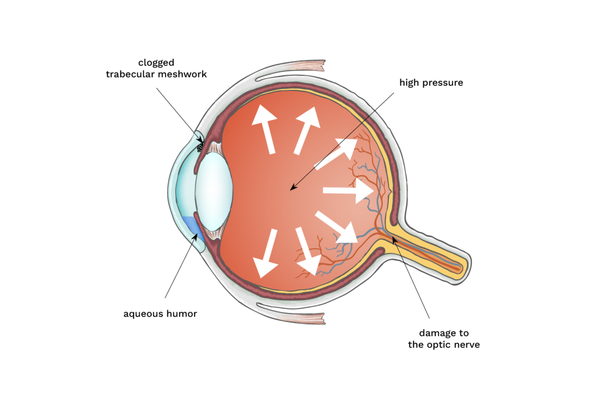
Glaucoma is a group of eye diseases that damage the optic nerve–the bundle of nerves that carries information from the eye to the brain. It can lead to vision loss and eventually blindness if left untreated. Continue reading for more information about the signs and symptoms.

Support Vision-Saving Research
More than 4 million Americans are living with glaucoma. Your gift can help fund research that could protect their sight and provide valuable information to the public.
What to Watch For
The most common types of glaucoma—open-angle and angle-closure—have completely different symptoms.
Open-Angle Glaucoma Symptoms
Most people who develop open-angle glaucoma, the most common form of the disease, don’t experience any noticeable symptoms at first. That’s why it’s critical to have regular eye exams so that your eye doctor can detect problems early on. Symptoms of open-angle glaucoma are:
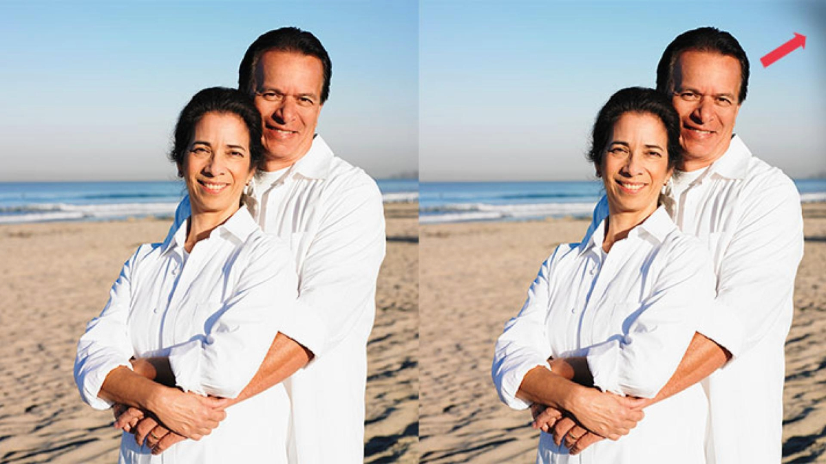
- Gradual loss of peripheral (side) vision, usually in both eyes
- Tunnel vision in the advanced stages
Acute Angle-Closure Glaucoma Symptoms
Acute angle-closure glaucoma is a medical emergency and must be treated immediately or blindness could result in one or two days. Signs and symptoms of acute angle-closure glaucoma include:
- Severe eye pain
- Nausea and vomiting (accompanying the severe eye pain)
- Sudden onset of visual disturbance, often in low light
- Blurred vision
- Halos around lights
- Reddening of the eye
Chronic Angle-Closure Glaucoma Symptoms
This type of glaucoma progresses slowly and can damage the optic nerve without symptoms, similar to open-angle glaucoma. Similarly, people with normal-tension glaucoma may not experience any symptoms until they begin to lose peripheral vision.
Neovascular Glaucoma Symptoms
Neovascular glaucoma is a secondary glaucoma where the eye angle is closed by “new” blood vessels, hence the name “neovascular.” It typically develops in eyes in which there is severe retinal vein blockage or severe diabetic eye disease. There are other causes too, including chronic retinal detachment, tumors, and ocular ischemic syndrome. Symptoms of neovascular glaucoma can include:
- Pain
- Redness
- Decreased vision

What is a Glaucoma Suspect?
A glaucoma suspect is someone who demonstrates one or more factors that put them at higher risk of developing glaucoma but does not yet have glaucoma damage. Sometimes, this is referred to as pre-glaucoma or borderline glaucoma.
You might be considered a glaucoma suspect if you have:
- High intraocular pressure or ocular hypertension
- Unusual or defective visual fields
- Other optic nerve features suggestive of glaucoma
Protect Your Vision
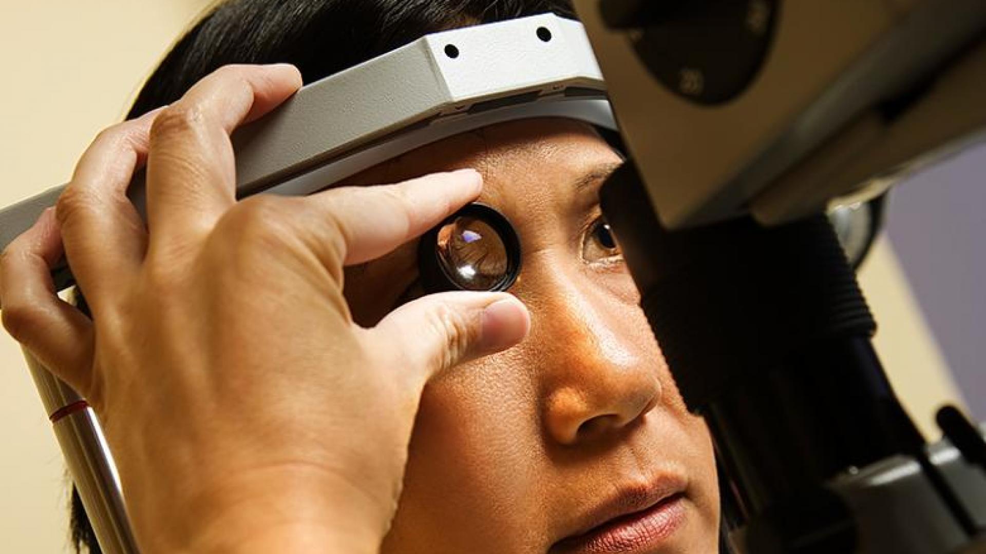
If you’re at high risk for glaucoma, you should have a dilated pupil eye examination at least every one to two years. Because glaucoma often has no early signs, regular eye exams are critical so that your eye doctor can detect problems early on. To help diagnose glaucoma, an ophthalmologist or optometrist will perform a comprehensive eye exam that may include the following tests:
Tonometry
This test measures the pressure inside the eye.
Pupil Dilation
Special drops temporarily enlarge the pupil so that the doctor can better view the inside of the eye.
Visual Field Testing
This test measures the entire area seen by the forward-looking eye to document straight-ahead (central) and side (peripheral) vision. It measures the dimmest light seen at each spot tested. Each time someone perceives a flash of light, he or she responds by pressing a button.
Visual Acuity Test
This test measures sight at various distances. While seated 20 feet from an eye chart, someonet reads standardized visual charts with each eye, with and without corrective lenses.
Pachymetry
The eye doctor uses an ultrasonic wave instrument to help determine the thickness of the cornea and better evaluate eye pressure.
Ophthalmoscopy
The doctor examines the interior of the eye by looking through the pupil with a special instrument. This test can help detect damage to the optic nerve caused by glaucoma.
Gonioscopy
The doctor uses this instrument to view the front part of the eye (anterior chamber) to determine if the iris is closer than normal to the back of the cornea. This test can help diagnose closed-angle glaucoma.
Optic Nerve Imaging
Imaging helps document optic nerve changes over time.
Search for a Glaucoma Clinical Trial
Clinical trials are crucial to advancing the most effective medical approaches. Today’s studies will lead to new standards of care in the future.

Resources
Recent Resources & Information

Expert Information
3 Subtle Signs Your Glaucoma May Be Getting Worse
In between doctor visits, you are the best advocate for detecting changes to your glaucoma. Discover the key symptoms to watch for and how early action can help protect your vision.

Glaucoma Chats
Can Glaucoma Be Prevented? The Science Behind Risk Reduction
Join us for a conversation with Dr. Rebecca Sarran, a leading glaucoma specialist, as she discusses the latest research on reducing the risk of glaucoma.

Expert Information
Ask An Expert: How Do I Know If I Have Glaucoma?
Protecting your eyes is one of the main actions you can take to prioritize your health as you age.

Expert Information
Understanding Secondary Glaucoma and Its Causes
Secondary glaucoma is a broad term that encompasses many types of glaucoma that are the result of certain medical conditions in the eye or the body.

Expert Information
Understanding Primary Angle-Closure Glaucoma: Symptoms, Diagnosis, and Treatment Options
Primary angle-closure glaucoma is the second most common form of glaucoma. Read about what it is, treatments, risk factors & more.

Expert Information
Open-Angle Glaucoma: Symptoms and Risk Factors
Learn why open-angle glaucoma rarely causes noticeable symptoms in the early stages of the disease, and why that is a problem. Learn who is at risk.


