


Macular Degeneration Research
Learn about an eye disorder that occurs more frequently in people who are very nearsighted.
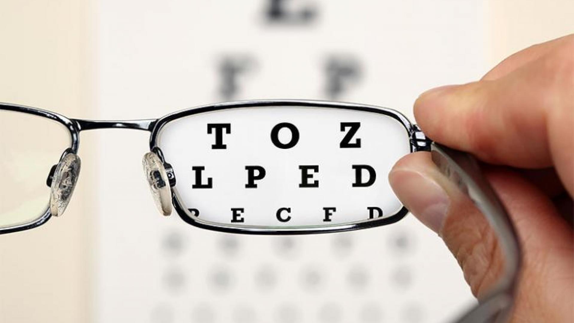
What is “High Myopia”?
Very nearsighted people are said to have “high myopia.” Myopia means nearsightedness, and the power of the lens that is needed to correct vision is measured in a unit called diopters. People who are very nearsighted, generally requiring glasses of -6 diopters or more, are at risk for myopic macular degeneration. The risk is higher as myopia becomes worse than -10 diopters. Myopic macular degeneration is distinct from age-related macular degeneration (AMD), although the two have some features in common. High myopia tends to run in families and is made worse by close-up work.
High Myopia and Degeneration of the Macula
High myopia is caused by having “long” eyes, with increased distance between the cornea in the front of the eye and the retina in the back. An increase in length of only a few millimeters can cause problems because the retina becomes “stretched.” Over time, this can cause cells in the center of the retina, the macula, to atrophy or die, causing a blind spot in the center of the visual field.

Learn more about the anatomy of the eye.
“Wet” Myopic Macular Degeneration
Some patients develop “wet” myopic macular degeneration, in which new blood vessels grow into the retina causing leakage of blood into the macula. This can cause sudden distortion of central vision, and, eventually, a blind spot in the central visual field.
Wet myopic macular degeneration can be treated the same way as wet AMD: with injections of anti-VEGF medications into the affected eye. VEGF is a protein made by the diseased retina that causes growth and leakage of abnormal blood vessels. It is often helpful to inhibit VEGF in diseases that involve leakage from new blood vessels that grow into the retina. Before the advent of VEGF inhibitors, about a decade ago, the abnormal vessels were treated with special lasers. Laser treatment is less common now.
High Myopia and Retinal Detachment
People with high myopia are also at risk for a retinal detachment, which is an emergency, since visual outcomes are better if the detachment is repaired sooner. The symptoms of a retinal detachment are: flashing lights in the peripheral vision, new floaters, and/or a “curtain” that blocks part of the visual field.
Summary
High myopia is a topic of current research. Hopefully better understanding of this condition will lead to improved preventions and treatments.
About BrightFocus Foundation
BrightFocus Foundation is a premier global nonprofit funder of research to defeat Alzheimer’s, macular degeneration, and glaucoma. Through its flagship research programs — Alzheimer’s Disease Research, Macular Degeneration Research, and National Glaucoma Research— the Foundation has awarded nearly $300 million in groundbreaking research funding over the past 51 years and shares the latest research findings, expert information, and resources to empower the millions impacted by these devastating diseases. Learn more at brightfocus.org.
Disclaimer: The information provided here is a public service of BrightFocus Foundation and is not intended to constitute medical advice. Please consult your physician for personalized medical, dietary, and/or exercise advice. Any medications or supplements should only be taken under medical supervision. BrightFocus Foundation does not endorse any medical products or therapies.
- Disease Biology
- Wet AMD
Resources
Related Articles and Information

Macular Chats
Geographic Atrophy: Making An Informed Treatment Choice
Learn about how to navigate treatment options, engage in shared decision-making with your healthcare team, and maximize your eye health.
Macular Chats
Wet AMD Treatments: Updates, Challenges, and Future Innovations
Join us for an in-depth discussion on the latest developments in wet age-related macular degeneration treatment.

Expert Information
Top 5 Expert Answers About Stargardt Disease
Explore key facts about Stargardt disease, a rare form of juvenile macular degeneration, with expert insights from a retina specialist.

Expert Information
Top Holiday Travel Tips for People with Low Vision
An expert shares must-know travel strategies and essential apps for a successful trip with low vision.