Glaucoma Imaging: Trends in Detection and Diagnosis
Featuring
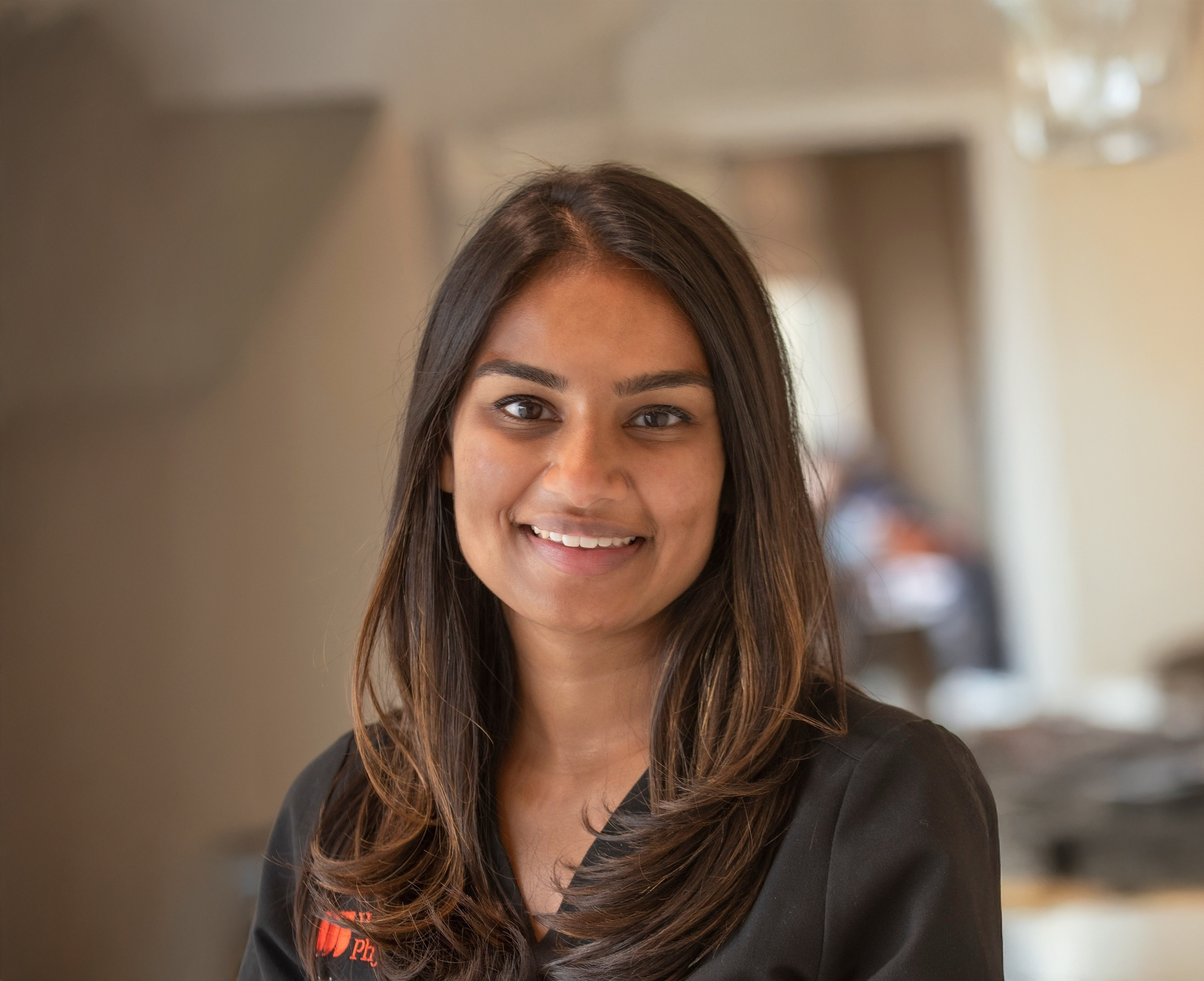
Sarah H. Van Tassel, MD
Associate Professor of Ophthalmology at Weill Cornell Medicine


Sarah H. Van Tassel, MD
Associate Professor of Ophthalmology at Weill Cornell Medicine

Optic nerve imaging techniques have undergone tremendous advancements over the last decade that will enable earlier glaucoma detection and diagnosis. Dr. Sarah Van Tassel outlined the current technology and took live questions from the audience.
Sarah H. Van Tassel, MD is Director of the Glaucoma Service and Glaucoma Fellowship at Weill Cornell Medicine Ophthalmology. Dr. Van Tassel specializes in glaucoma evaluation and treatment, including medical management, laser, and surgical procedures. She also performs cataract surgery and is up-to-date on the latest advances in microinvasive glaucoma surgery (MIGS). A native of Missouri, Dr. Van Tassel completed her residency in ophthalmology at Weill Cornell Medical College and her fellowship in glaucoma at Duke University. Dr. Van Tassel’s interests include personalized glaucoma care, surgical outcomes, ophthalmic imaging, and the intersection of mental health and glaucoma.
MS. KACI BAEZ: Hello, and welcome to today’s BrightFocus Glaucoma Chat. My name is Kaci Baez, and I’m VP of Integrated Marketing and Communications at BrightFocus. I’m excited to be here with you today as we talk about “Glaucoma Imaging: Trends in Detection and Diagnosis.” Our Glaucoma Chats are a monthly program, in partnership with the American Glaucoma Society, designed to provide people living with glaucoma and the family and friends who support them with information straight from the experts. BrightFocus Foundation is committed to investing in bold research worldwide that generates novel approaches, diagnostic tools, and life-enhancing treatments that serve all populations in that fight against age-related brain and vision diseases. Now I would like to introduce today’s guest expert. Dr. Sarah Van Tassel is Director of the Glaucoma Service and Glaucoma Fellowship at Weill Cornell Medicine Ophthalmology. Dr. Van Tassel specializes in glaucoma evaluation and treatment, including medical management, laser, and surgical procedures. She also performs cataract surgery and is up to date on the latest advances in microinvasive glaucoma surgery. A native of Missouri, Dr. Van Tassel completed her residency in ophthalmology at Weill Cornell Medical College and her fellowship in glaucoma at Duke University. Her interests include personalized glaucoma care, surgical outcomes, ophthalmic imaging, and the intersection of mental health and glaucoma. Welcome. We’re so excited to have you here today, Dr. Van Tassel.
DR. SARAH VAN TASSEL: Thank you for having me. I’m delighted to be here.
MS. KACI BAEZ: Awesome. Our first question that we have is: Can you start us off by just explaining how people usually get diagnosed with glaucoma, maybe for those who aren’t familiar? Just as a reminder, because sometimes there are new diagnoses and advancements and such. How is the disease typically detected?
DR. SARAH VAN TASSEL: Yeah, let me just start by briefly saying I know our audience knows a lot about glaucoma, but what is glaucoma? It’s damage to the optic nerve, and the optic nerve is the electric cable that sends the signal of vision from the back of the eye to the brain. And in order to see well, you need that electrical conduit to be working perfectly. The way that people most commonly get diagnosed with glaucoma is because of abnormalities that are seen on an exam of the optic nerve. So, what are those scenarios? I would say the most common scenario in my practice is that someone is referred for glaucoma because they underwent routine eye care in the community and the optometrist or ophthalmologist that they saw observed the possibility of glaucoma. Usually, that’s because someone is going for glasses or contact lenses for blurry vision and then this gets picked up on exam. I would say the second scenario is that someone knows that they might be at risk for glaucoma because they have a first-degree relative with glaucoma, so they seek out the care of a glaucoma specialist because they’re particularly interested in being vigilant. And I would say the third scenario, but far and away the least common, is that someone has symptoms from glaucoma and then they come in to see me because they’re complaining of peripheral vision loss or decreased vision.
MS. KACI BAEZ: Thank you. Does diagnosis and detection vary by type of glaucoma or stage of disease?
DR. SARAH VAN TASSEL: Yeah, it can. When someone comes in for a glaucoma evaluation, they typically get the same battery of tests, regardless of the type of glaucoma or the stage of glaucoma. What is that? It’s often very first a peripheral vision test and then often gonioscopy, which is using a special contact lens that sits on the surface of the eye to look at the natural drain inside the eye to tell if the drain is open or closed, because that distinguishes open angle glaucoma from narrow angle or closed angle glaucoma, and the treatments can be different. And then depending on the scenario, the patient likely gets their pupils dilated with dilating eye drops, and then they would get imaging of the optic nerve, which might be either a photograph—just like a digital photograph of the optic nerve and retina—and in 2024, it’s even more likely to be an OCT, which is sort of the heat map of the optic nerve and retina that we’re going to speak more about.
And even though everybody gets that same testing, it is often the case that in very early glaucoma, the OCT is a bit more helpful because in early glaucoma, the peripheral vision is, by definition, normal, so the peripheral vision test tends to provide less information, but we can detect the subtleties of damage to the optic nerve by looking at the OCT. On the opposite end of the spectrum is the scenario where a patient has really far advanced glaucoma, and in that situation, the optic nerve might already be so thin or so damaged that it is hard to pick up signs of change on the OCT—we can tell that the optic nerve is abnormal, but we can’t detect further abnormality over time. In those patients, we really rely on the peripheral vision test because that tells us what the patient is experiencing and helps us to look for subtle signs of change. For people in the middle, in the moderate to maybe early severe category, very often the visual field test and the OCT can provide meaningful information that we’re keeping track of very closely to look for signs of change. All of our interests in glaucoma are aligned to look for change—that’s kind of the name of the game. Even if the peripheral vision or the OCT are abnormal, as long as they are stably abnormal, then we’re sort of winning, so to speak; we’re doing our job of preventing progression. It’s when things are getting worse that we feel like the disease is winning.
MS. KACI BAEZ: Now, you referenced OCT and optic nerve imaging, so could you explain a little more about what that is, what you’re looking for, and how do you know if the disease is winning, as you said?
DR. SARAH VAN TASSEL: Yeah, so OCT is essentially like a heat map, in a sense. It’s using beams of light to measure the thickness of the various parts of the eye tissue. And there are two things we care the most about when we’re watching for glaucoma, and that tends to be the thickness of the retina in the area right around the optic nerve, and the term for that is retinal nerve fiber layer, RNFL. And that part of the tissue essentially contains the little tails that communicate the information from the photoreceptors, which is kind of a common colloquial term, although there are other cells in the retina, but essentially the retinal nerve fiber layer transmits the visual signal from the photoreceptors into the optic nerve and along the brain. We want to measure the thickness of those tails because if that area—the RNFL—is getting thinner over time, that’s a sign of glaucoma. And so, the OCT makes a heat map of that issue, and if you’ve seen it on the screen or on the printout in your physician’s office, then you know that it is very often green and yellow and red for good and medium and thin—very thin.
And then increasingly, ophthalmologists are very interested in also measuring the macula. Very often, we think of the macula as the domain of retina specialists, but we know that the retinal ganglion cells, which live in the macula, are the ones that are actually being damaged in glaucoma. So, rather than only measuring the thickness of those tails as they communicate the information into the optic nerve, we can also measure the thickness of those cell bodies in the macula, the back of the retina, essentially. And that can also provide helpful information about glaucoma, and we can detect specific patterns of abnormal cells that clue us in to whether someone has glaucoma and also clue us in to whether someone is progressing. And I think what I was referring to earlier—winning—is just that we always want to make sure that these tests are stable from visit to visit. And if they’re not stable from visit to visit, we have to decide: Is there a reason for that that isn’t glaucoma? Is something else happening in the eye, or is the glaucoma getting worse? And then, the name of the game is lowering the pressure further. There are many, many things that have been explored for glaucoma—nutritionally and lifestyle intervention and other medications—but we know that right now, the best evidence tells us that lowering the pressure inside the eye is the only way to prevent glaucoma from getting worse. And sometimes we lower the pressure, and the patient’s glaucoma is still getting worse, and so we have to lower the pressure even further with more laser or more surgery or more eye drops. And so, the decision about whether to pursue additional treatments and additional interventions is based on these data that we’re collecting during the office visit.
MS. KACI BAEZ: How is the optic nerve imaging different from the retinal imaging testing, which is the testing that a retina specialist does?
DR. SARAH VAN TASSEL: Yeah, that’s a great question, because it’s very commonly the case in my practice, which has 30 or so doctors of all different specialties, that a patient will say, “Well, I just have these pictures taken last week.” And I say, “Oh, I know the experience is so similar, but the information is actually so different.” Very commonly, the retina specialist also takes a photo, just a digital-type photo as glaucoma specialists sometimes takes, but then they also take OCT. It’s based on this same technology principles, but the anatomical tissues inside the eye that they are looking at and the measurements they’re taking are different than what we’re looking at as glaucoma specialists. And so, the retina specialists are also using OCT of the retina, but they’re not measuring the thickness of the ganglion cells, which is what the glaucoma specialists want to know, and instead they’re looking for things like wet and dry age-related macular degeneration or swelling from diabetes in the retina and the macula or evidence of microscopic strokes in the back of the eye—things like that. So, the technology is the same, but the way the information is distilled on the printout and whatnot is actually quite different. So, for patients who have a reason to see both a retina specialist and a glaucoma specialist, you really do need the testing for both visits.
MS. KACI BAEZ: That’s great information. Thank you. And so, how is retinal imaging testing different and the OCT different from the visual field test, which you also referenced?
DR. SARAH VAN TASSEL: Right. So, the visual field test, in brief, for people who perhaps haven’t taken it, one eye is covered, and you take the test with the other eye. And essentially, the person taking the test—the patient—looks at a center target and tries to only look at that target without looking around. And then in the peripheral part of the vision, little lights kind of twinkle or blink, and without looking at them, the patient then clicks a button to say that they saw the stimulus in that periphery. It’s a really hard test to take because your brain wants to look at that peripheral light, but the machine knows. There are some metrics that tell us when you’re looking around. And it’s really only a helpful valid test if the center light is looked at the whole time and then the peripheral vision is tested. And patients tell us that the test—the visual field test, that is—is really hard to take, and that it’s very frustrating to take, and those are true statements. But it is so important because of the tests that we do in glaucoma. It’s the only one that really tells us what the patient is experiencing from their glaucoma. And there’s no other way with imaging or with photos to understand the actual function of the visual system. So, we want to know not just how the cells look in the pictures and how the tissue looks in the pictures, we want to know how it is working for the patient and what they are seeing. And the visual field tells us that, and it’s important, and there are some advances in visual field that will hopefully make it easier for patients and less burdensome—wearing a headset on your head, for example, rather than doing it in the big machine at the office. But it’s certainly different. It tests different information, it provides different information, and it’s really meaningful for glaucoma surveillance.
MS. KACI BAEZ: That’s great. It seems like we are so fortunate to have these advancements and to even have these tests available. What other tests are important when it comes to detecting and diagnosing glaucoma?
DR. SARAH VAN TASSEL: I mentioned it in brief just a few minutes ago, but I would say one of the tests that’s really important is gonioscopy. That’s the test I was describing where a glass contact lens is placed on the surface of the patient’s eye and then allows the physician to see the natural drain inside the eye. This is really the primary way to distinguish open angle glaucomas from closed angle glaucoma.
And it matters a lot because the treatment for those glaucomas is different, and I would say I see people in my office who have been followed elsewhere for many, many years, and they say pretty confidently that they’re positive they’ve never had that test done before when I do it. And it really is so important because it provides really helpful information about the type of glaucoma, and then it opens doors for treatments like laser therapies that the literature tells us can be really even better than eye drops for some types of glaucoma. So, I would say gonioscopy is another test that’s important.
Central corneal thickness, or CCT, is an important number—that’s a measurement of the thickness of the clear part of the eye. That’s a one-time test for most people. So, if you’ve had it done even once at your doctor’s office, that’s enough because that’s not a number that changes over time. But we know that the corneal thickness changes the patient’s pressure measurement a little bit. People with thick corneas tend to measure a little higher on their eye pressure, and conversely, thin corneas tend to measure a little lower on the eye pressure. And that kind of makes sense if you think about putting the same pressure using like a bike wheel pump into a balloon or into a basketball, you know that even if the pressure is the same in both, the balloon is going to feel softer than the basketball because the basketball has that thicker wall. And you do get some measurement error in the pressure, which is why central corneal thickness matters. And then the other thing we know is that people with thick corneas just tend to be a little bit less at risk for glaucoma. So, it tells us some information about the tissues of the eye that’s helpful. And then I would say, of course, the other test, which we haven’t talked as much about but is implicit in every glaucoma visit, is a pressure check. So, that always gets done, and we call that, of course, the IOP—intraocular pressure.
MS. KACI BAEZ: Great. And how long does imaging take, and how often should it be done?
DR. SARAH VAN TASSEL: I would say it takes longer than you think it ought to. That’s the short answer to that question because there are a lot of touch points during the visit at an ophthalmologist office or a glaucoma specialist office. You might get the visual field test with one technician, and then you wait for a different technician to see you for things like the vision and the pressure and maybe a glasses check, and then your pupils get dilated, and then you might wait for yet a third person to come and take your images, and then you’re waiting to see the glaucoma specialist. So, it can take well in excess of an hour and a half, and that’s definitely a source of frustration for patients and really kind of a ripe area for innovation, as well. A lot of doctors and a lot of companies are working really actively to think about what tests can be done at home and what tests are really necessary and how we can improve office efficiency, because so many people need glaucoma care. But I would say, on a best day, where everything is moving efficiently, it is impossible in my office to get those tests in less than about an hour and 20 minutes.
And then, the question about how often should they be done: It really depends on the severity of the patient’s glaucoma and the physician’s impression of whether or not the glaucoma is progressing. I typically tell patients that if you have severe glaucoma, you’ll be seen every 3–4 months, and we might get a visual field and/or an OCT at each of those visits. Patients with moderate glaucoma, I typically see in the 4- to 6-month range, and then patients with mild glaucoma or glaucoma suspects under evaluation for glaucoma, I often see once a year. But if you’ve been seen in that pattern and then all of a sudden it seemed like things were progressing, your doctor might want to see you more often to reestablish the tempo of what’s happening. Sometimes longer periods between visits make it harder to detect change, so at least once a year; in many cases, more often.
MS. KACI BAEZ: Are there any risks to these tests? And what are the benefits?
DR. SARAH VAN TASSEL: Yeah. The benefits, that’s the easiest to discuss. Glaucoma, by definition, in most people is an asymptomatic disease. You don’t know you have glaucoma for a really long time, which is why people ultimately can go blind from glaucoma—because it can progress for so, so long without detection, and then if you wait for symptoms to develop, it’s quite advanced and can be hard to treat.
And that’s just because the two eyes work so well together and the brain is so smart, and the brain just puts the visual signal that it’s getting about the world together and sort of tricks you into thinking you’re seeing more than you might be. And so, the clear benefit of the testing is detecting glaucoma and detecting it as early as possible so that patients can get the therapies that they need. The risks of testing are small, and we’ve always talked about time and frustration, but those are risks, and those are able to be overcome. I think the elephant in the room for all medical testing, perhaps, is money. Sometimes it can be difficult for a patient to know in advance what their bill for a visit might be. Much to many patients’ surprise, it’s actually very hard for the doctors in the doctor’s offices sometimes to know what that’s going to look like also, and that is a big topic beyond the scope of today’s discussion. But the unknowns about the bill are a risk, but I would say there’s really no harm to your eyes or to your person for the testing.
MS. KACI BAEZ: Thank you. After testing, patients will typically get a report from their doctor or in a patient portal, and if their result said, “abnormal”—and I think this is true for any test, especially when you get the results before you have a chance to talk to your doctor, you can start to panic—can you comment on what those reports might mean and maybe the best way for somebody to work with their doctor to understand them?
DR. SARAH VAN TASSEL: Yeah. Well, I think it’s such a tough balance because I think that the move toward information being more available and more accessible to patients and in a really timely fashion has just extraordinary benefits. But I think the challenge is that the things that doctors write in the chart are by and large not intended for patients—they’re intended for other physicians or for themselves. Notes to yourself that you write for a future visit to keep track of things or notes that you write to other providers of care on a patient’s team or documentation that you write to satisfy billing requirements for the testing.
And so, very often, it does say abnormal because, by definition, if someone’s under evaluation for glaucoma, their testing probably is, at least in part, abnormal. But what we worry about isn’t so much the abnormal because glaucoma is damage that can’t be reversed, but what we are looking for, as I’ve discussed, is change over time. So, what we want is for the abnormal scan to be stable. And generally speaking, if there is a change, then at that visit your physician would have discussed with you concerns about change and the decision process for whether the therapies should be changed or escalated in some way. But I would say that I write “abnormal” on the bulk of my OCT interpretations, so that in and of itself can be disheartening to patients but shouldn’t be a reason for concern.
MS. KACI BAEZ: Right. We’ve touched on optic nerve imaging. Is that only used for diagnosis, or can imaging—such as the optic nerve imaging or possibly other imaging—predict or detect progression following a diagnosis?
DR. SARAH VAN TASSEL: Yeah, those are the two things that we use it for—both to make the diagnosis itself and then to monitor for change over time. And so, there’s what we call intraday variability. If you take the scan even on a healthy eye today and you take it again tomorrow, because we’re measuring in microns, there’s going to be a different number generated for each of the measurements, and so you need several tests over time to really understand what the average looks like for that patient and then to detect whether something that looks different or changed, is that really change or is it just intraday variation? We very much use this same panel of testing for making the diagnosis itself and then for monitoring for treatment … for progression. I guess we’re saying that that’s the reason that follow-up appointments are so important, because the literature tells us that the biggest risk factor for losing vision to glaucoma beyond having advanced glaucoma is just getting lost to follow-up because you don’t know whether things are stable. The only way to know for sure that they’re stable is to repeat the testing at your doctor’s office. And so, we know that nothing really matters more in terms of vision preservation from glaucoma than seeing your ophthalmologist at the interval that’s been recommended.
MS. KACI BAEZ: Absolutely, it’s always so important to keep your appointments and to also set reminders for yourself, I think, especially when you have so many appointments and there’s so many different steps involved and maybe different specialists and a team of providers who have to work together. It actually takes a lot of work, I think, to take care of oneself. And when you have a disease like glaucoma, that’s a lot. That’s not just, “Go to the doctor once a year”; it really takes a commitment to take care of your eye health. And so, just how have imaging techniques advanced over the years?
DR. SARAH VAN TASSEL: Well, the advancements in OCT have really been leaps. As recently as 20 or so years ago, really, ophthalmologists were relying primarily on the photos themselves and their clinical exam of the optic nerve rather than on the OCT. And the advancements in OCT have been in how quickly the image is captured while the patient is looking at the camera, so to speak, and then on the fidelity of the testing, the resolution in terms of the microns—much the same way that the digital cameras we used to buy had 3 megapixels, for example, and now, they all have an excess of 15 or so. The resolution of the OCT and the fidelity of the testing has improved tremendously. And in the visual fields, there’s probably been a little bit less advancement, but as I mentioned earlier, their headsets—and we call them virtual visual fields—are really a hot, hot topic where we’re getting ready to see really, really rapid change over time. And I think in my practice lifetime, we’ll probably be able to capture OCTs in patients’ homes or with their smartphones, for example. So, we haven’t yet arrived in terms of advancement.
MS. KACI BAEZ: Thank you. We know that imaging is very helpful, especially early in glaucoma for diagnosis. For those who are already diagnosed, what message would you pass on for their family members?
DR. SARAH VAN TASSEL: I think the most important gift you can give your family members is just to tell them about your diagnosis or even the suspicion for glaucoma and encourage them to have an eye exam also. I think it’s so interesting because so many people say, “Oh, in my culture, we don’t really talk about our health,” and “In my culture, we don’t talk about our health.” And I will tell you, in New York City, I see a lot of cultures, and everybody thinks their culture doesn’t talk about their health. But I think it’s up to the next generation of families to try to break down some of those barriers and to be a bit more transparent about health at big family gatherings or around holidays and whatnot just to check in and see what you might be at risk for. But importantly, if you have a first-degree relative with glaucoma, you should be tested. And if you are that first-degree relative, you should avail your family members of the opportunity to know that they should be screened. There’s a growing role, probably, for genetic testing in this space also. But right now, in 2024, the most important thing you can do is just to have your family members be examined by an ophthalmologist.
MS. KACI BAEZ: That’s a great point of people not talking about their health. It does seem like a generational challenge rather than cultural. It just seems like there’s been a shift to people in the younger generations who are more willing to talk about their health, and it’s shifting in the right direction. We did get a listener question, and it’s: The eye pressure test tests the pressure in the front of the eye. Since the front of the eye is separated from the back by the lens, do we know the pressure in the back of the eye?
DR. SARAH VAN TASSEL: Thanks for that good question, listener. There’s some complexity to that question, but generally speaking, inside the wall of the eye, the pressure is very similar. There are actually some new instruments and things that are being investigated for really measuring the inside of the eye pressure, implants that go inside the eye that fit in various spaces that will be able to generate real-time data to a smartphone or to another device about what the pressure is inside the eye. And none of those are ready for prime time, but I would say that the pressure inside the eye is similar throughout the various chambers, although there are some differences that the listener observed. But I would say that, importantly, it’s not even about getting the number perfectly because you can get glaucoma at any number pressure—35 or so percent of patients have never had high pressure. But what matters is that measuring from the outside of the eye is a proxy for what’s going on inside the eye. And so, because we’re monitoring for change and because we’re monitoring for pressure lowering from whatever the patient’s baseline was, we know that even measuring from outside the eye, we’re able to detect change. That’s a good question.
MS. KACI BAEZ: Outside looking in.
DR. SARAH VAN TASSEL: Yeah.
MS. KACI BAEZ: During our prep call you gave an analogy comparing images of the eye to a still image of race cars at the Indy 500, and I wondered if we could maybe wrap up our call today with you sharing that with our listeners.
DR. SARAH VAN TASSEL: Yeah, I’d love that. So, I tell patients, and I didn’t come up with this analogy, though. I don’t know who to credit. But sometimes I meet a patient for the very first time and I’m looking at their testing and maybe they have some peripheral vision loss and they have some abnormalities of their OCT, and they say, “Well hey, doc, is it getting worse?” And I say that looking at this testing is like looking at that photograph from the Indy 500. I can tell the car is moving, but I don’t know how fast it’s going, and that’s often the case with glaucoma. When you meet a new doctor for the first time, they can tell that you have glaucoma, but unless you follow up routinely and get the recommended surveillance testing, we don’t really know how quickly it’s advancing, and that just speaks to the reason why testing and frequent testing is so, so, so important for catching this disease as early as possible and then treating it appropriately, aggressively.
MS. KACI BAEZ: Absolutely. Set your reminders to your tests, listeners. And thank you Dr. Van Tassel for all of the important information you shared with us today. Next month, on May 8, we will dive into a hot topic—”Exploring Artificial Intelligence in Glaucoma: Challenges, Opportunities, and Hope.” We hope that you will join us again on May 8, and thanks so much for tuning in today.
BrightFocus Foundation is a premier global nonprofit funder of research to defeat Alzheimer’s, macular degeneration, and glaucoma. Through its flagship research programs — Alzheimer’s Disease Research, Macular Degeneration Research, and National Glaucoma Research— the Foundation has awarded nearly $300 million in groundbreaking research funding over the past 51 years and shares the latest research findings, expert information, and resources to empower the millions impacted by these devastating diseases. Learn more at brightfocus.org.
Disclaimer: The information provided here is a public service of BrightFocus Foundation and is not intended to constitute medical advice. Please consult your physician for personalized medical, dietary, and/or exercise advice. Any medications or supplements should only be taken under medical supervision. BrightFocus Foundation does not endorse any medical products or therapies.

Can lifestyle changes help reduce glaucoma risk? Join us as glaucoma specialist Jullia A. Rosdahl, MD, PhD, discusses how diet, exercise, sleep, and other non-drug interventions may impact eye pressure and overall eye health. Learn about the latest research on nutrition, supplements, and daily habits that could support vision preservation.

Join Dr. Lawrence S. Geyman as he outlines effective ways to find support for your glaucoma diagnosis.

Learn about the similarities and differences between these two vision diseases, whether someone can develop both conditions at the same time, and how to reduce your risk.

Join us for an insightful conversation with Dr. Jaehong Han about the different types of glaucoma, including open-angle, angle-closure, normal-tension, and more, discussing who is most affected by each and how treatments can vary.
