Breakthrough Discovery Reveals New Pathway to Lowering Eye Pressure
The findings from this first-of-its-kind study can help improve current glaucoma drugs and lead to the development of new treatments.


The findings from this first-of-its-kind study can help improve current glaucoma drugs and lead to the development of new treatments.

A tiny part of the eye could play an oversized role in lowering eye pressure.
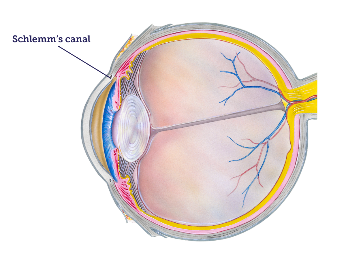
By studying cell activity in Schlemm’s canal, a minuscule part of the eye’s drainage system, BrightFocus-funded researchers have identified a potential new pathway for improving and developing drugs to treat glaucoma.
First recognized in 1830 by German anatomy scientist Friedrich Schlemm, Schlemm’s canal is a unique, ring-shaped vessel that circles the cornea. It, along with the trabecular meshwork, acts as a gatekeeper to maintain the correct amount of fluid in the eyes and regulate the eye pressure.
The team, which included National Glaucoma Research grantees Myoung Sup Sim, PhD, and Paloma B. Liton, PhD, of Duke University, found that the activity of the cells within Schlemm’s canal could lead to an important means of lowering eye pressure. They reported the results of their study in the July 2023 issue of the journal Autophagy Reports.
Glaucoma is a group of eye diseases in which the optic nerve at the back of the eye becomes damaged from too much pressure within the eye. In most cases, this damage is related to increased eye pressure caused by a buildup of fluid called aqueous humor.
The aqueous humor is the clear fluid in the front of the eyeball, between the lens and the cornea. It plays a pivotal role in nourishing the eyeball and helping to maintain its shape. It’s different from tears since aqueous humor is inside the eye.
In healthy eyes, there is a balance between the fluid that is made in the eye and the fluid that leaves the eye. In glaucoma, however, the outflow of aqueous humor becomes blocked. When the fluid has nowhere to go, it builds up in the eye, creating high pressure that can harm the optic nerve. Learn more about the importance of the eye’s drainage system in glaucoma.
Symptoms of glaucoma can start so slowly that they often are not noticed at first. A dilated eye exam is the only way to diagnose the disease. There is no cure for glaucoma, but early treatment can often stop the damage and protect vision.
To leave the eye, excess aqueous humor flows through spongy tissue called the trabecular meshwork and into Schlemm’s canal. For many years, trabecular meshwork has been the focus of studies to manage eye pressure. The researchers, however, found that Schlemm’s canal could be more important.
This first-of-its-kind research study identified a new way in which autophagy is activated in the canal. Autophagy is the process in which cells absorb other cells, reusing their old or damaged parts to help the cells work more efficiently. It is emerging as a critical pathway to treating several diseases, including glaucoma.
The findings from this unique study can help advance and improve current glaucoma drugs. In addition, the findings provide a more complete understanding of how autophagy regulates eye pressure and could lead to the development of new drugs to treat glaucoma more efficiently and safely. Future studies will focus on the molecular mechanisms involved in autophagy within Schlemm’s canal.
BrightFocus Foundation is a premier global nonprofit funder of research to defeat Alzheimer’s, macular degeneration, and glaucoma. Through its flagship research programs — Alzheimer’s Disease Research, Macular Degeneration Research, and National Glaucoma Research— the Foundation has awarded nearly $300 million in groundbreaking research funding over the past 51 years and shares the latest research findings, expert information, and resources to empower the millions impacted by these devastating diseases. Learn more at brightfocus.org.
Disclaimer: The information provided here is a public service of BrightFocus Foundation and is not intended to constitute medical advice. Please consult your physician for personalized medical, dietary, and/or exercise advice. Any medications or supplements should only be taken under medical supervision. BrightFocus Foundation does not endorse any medical products or therapies.

Duke University School of Medicine

Duke University Eye Center
