Simultaneous Structural and Functional Imaging of the Retinal Pigment Epithelium

Principal Investigator
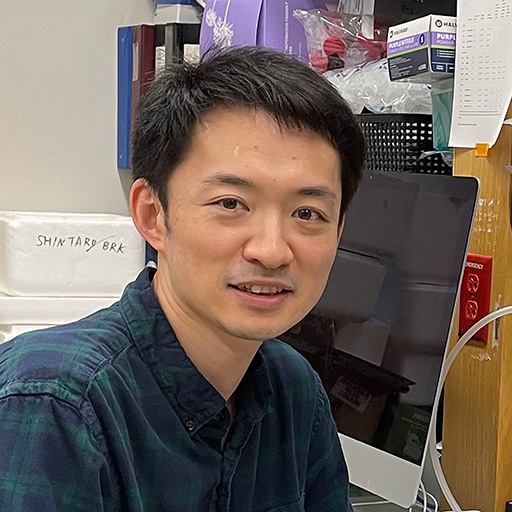
Omid Masihzadeh, PhD
University of Colorado School of Medicine
Aurora, CO, USA
About the Research Project
Program
Award Type
Standard
Award Amount
$120,000
Active Dates
July 01, 2014 - March 01, 2017
Grant ID
M2014014
Co-Principal Investigator(s)
David Ammar, PhD, University of Colorado Denver
Goals
The progression of age-related macular degeneration (AMD) coincides with structural changes to the eye. Currently, clinicians and researchers use visible light and histological sectioning to study these changes but are limited to detections of only fairly large structures and lack the necessary information associated with the disease. We propose a new paradigm in light microscopy to detect both changes to the eye’s structure as well as functional abnormalities associated with AMD. Currently, this detection method is unknown and unavailable to researchers and clinicians. We propose to investigate the viability of this technique for studying AMD.
Summary
In this proposal, we investigate the possibility of vivo imaging studies for age-related macular degeneration (AMD) and ocular diseases in general. While many of the pathognomonic changes of AMD, such as drusen and alterations in retinal pigment epithelium (RPE) physiology, are documented by traditional light or fluorescent microscopy, the molecular mechanism that causes disease onset remains unclear and is best illuminated through longitudinal studies of live animal disease models. Therefore, there is a vital need for dynamic monitoring of living cellular processes, so-called functional imaging, at the subcellular level that would allow for real-time monitoring of the initiation and progression of AMD. We hypothesize that coherent anti-Stokes Raman spectroscopy (CARS), a chemically specific multiphoton spectroscopic technique, can track the onset and progression of AMD by monitoring drusen formation in intact eyes.
Important functional information, such as metabolic states or molecular interactions that promote AMD pathology, can only be studied in vivo. Compared to currently used clinical and nonclinical imaging, CARS microscopy is a relatively young technology. Nonetheless, due to its inherent capability for in vivo diagnostics, its potential impact is enormous. A natural progression from our study, if successful, would be the construction of economically viable systems for studying the pathognomonic changes of AMD in live animals using the CARS modality.
Mice that lack the anti-oxidant enzyme Sod1 develop drusen, thickened Bruch’s membrane, and choroidal neovascularization characteristic of AMD. Here, we will examine our hypothesis that CARS can be used to non-invasively and non-destructively study the onset and progression of AMD in Sod1-/- mice.
Grants
Related Grants
Macular Degeneration Research
The Novel Role of an Intracellular Nuclear Receptor in AMD Pathogenesis
Active Dates
July 01, 2024 - June 30, 2026

Principal Investigator
Neetu Kushwah, PhD
Current Organization
Boston Children’s Hospital
Macular Degeneration Research
Exploring How NRF2 Protein Reduces RPE Cell Damage by Cigarette Smoke
Active Dates
July 01, 2024 - June 30, 2026

Principal Investigator
Krishna Singh, PhD
Current Organization
Johns Hopkins University School of Medicine
Macular Degeneration Research
The Development of a Transplant-Independent Therapy for RPE Dysfunction
Active Dates
July 01, 2024 - June 30, 2026

Principal Investigator
Shintaro Shirahama, MD, PhD
Current Organization
Schepens Eye Research Institute of Massachusetts Eye and Ear


